Gray Appearance After Dental Implants: Causes & Fixes
You invested time, money, and hope into a dental implant. You expected a seamless, radiant smile. Instead, you notice a faint gray shadow. The gum around your new implant looks dusky, bluish, or metallic. The tooth itself might seem darker than its neighbors. Your first thought is often panic: “Is the implant dying?” or “Did I make a mistake?” A gray appearance after dental implant treatment is a known aesthetic complication. It does not necessarily signal a failing implant, but it does indicate an underlying issue that needs identification and correction. This article will explore every reason why an implant can look gray, from the bone deep below to the crown on top. We will provide clear, realistic solutions for each scenario. Your goal is an undetectable restoration. Understanding the source of the gray is the first step.
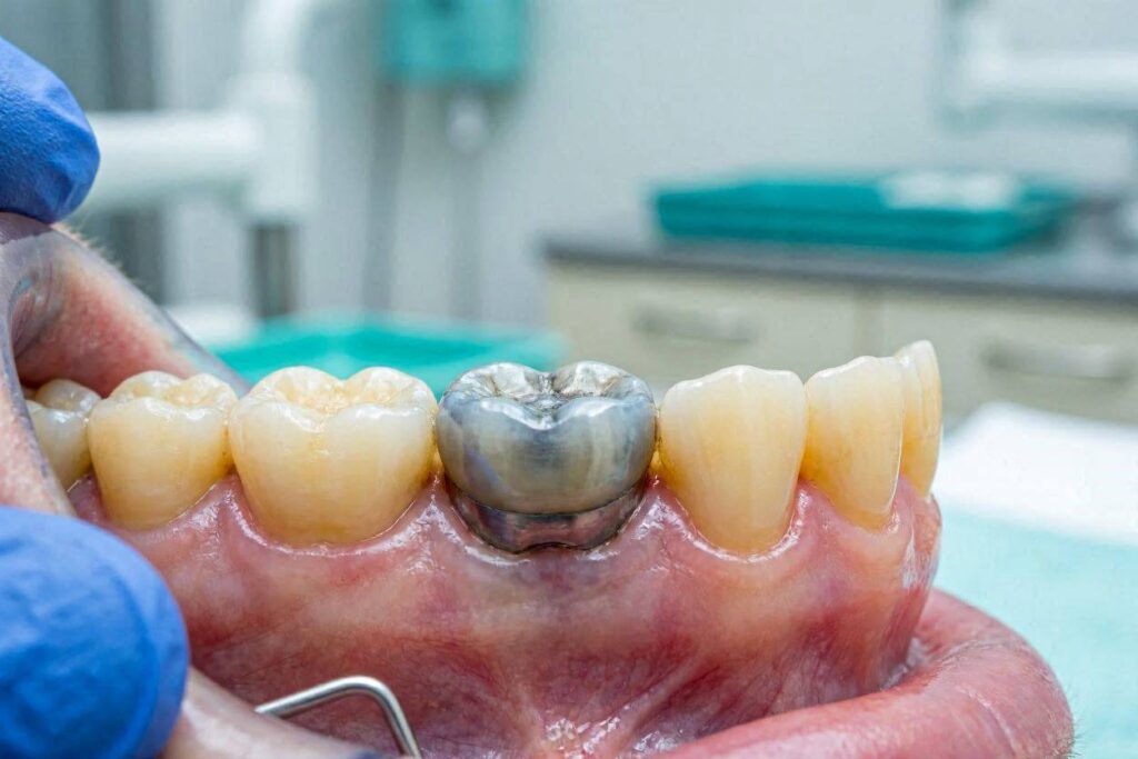
The Optical Anatomy of a Natural Tooth vs. an Implant
To understand the gray, you must first appreciate the optical complexity of a natural tooth. A tooth is not a solid white block. It is a translucent, layered structure. The outer enamel is semi-translucent, like frosted glass. It transmits light and reveals the color of the underlying dentin. Dentin is a warm, yellowish-bone colored tissue. This combination of translucent enamel over yellow dentin gives a vital, vibrant appearance. Light enters the enamel, reflects off the dentin, and bounces back out. The root of the tooth is covered by a thin layer of cementum and then by gum tissue and bone. The gum and bone have their own color—a healthy, vascular pink. Light diffuses through the gum tissue. The tooth root helps block light, but a natural root, being yellowish dentin, does not create a dark shadow.
The Implant Components and Their Colors
An implant is an assembly of manufactured materials with very different optical properties.
- The implant post, buried in bone, is typically made of commercially pure titanium or a titanium alloy. Titanium is a dark, metallic gray. It is completely opaque.
- The abutment connects the post to the crown. It can be made of titanium (gray), gold-shaded titanium (gold), or zirconia (white).
- The crown is the visible tooth. It is made of a ceramic material like porcelain fused to metal (PFM), or all-ceramic like lithium disilicate (e.max) or zirconia.
The gray appearance can originate from any of these layers. The light must pass through the gum, the abutment, and the crown. If any component blocks, absorbs, or scatters light in an unnatural way, a gray effect is the result.
The Primary Causes of a Gray Appearance
A systematic analysis is essential. The gray can come from the tissue, the implant components, or both. Let’s categorize the root causes.
1. Peri-Implant Soft Tissue Discoloration
This is the most common cause of a gray appearance when the patient sees a dark, bluish or purplish hue in the gum itself, not necessarily in the crown. The gum tissue is the lens through which we view the underlying structures.
Thin Biotype and Titanium Show-Through
Every person has a genetically determined gum biotype. A thin, scalloped biotype is characterized by a delicate, almost translucent gum. The underlying blood vessels and, critically, the underlying titanium implant and abutment can visually influence the tissue color. The dark metallic gray of the titanium absorbs and blocks light, creating a zone of shadow under the gum. When light hits the gum, it transilluminates the thin tissue and reveals this darkness. This is not a disease. The tissue is healthy. It’s akin to a dark hair root showing through fair skin. This is a true optical phenomenon, present from the day the abutment is placed and often stable over time unless recession occurs, which worsens it.
Peri-Implant Mucositis and Peri-Implantitis
This is a disease state. Inflammation changes tissue color dramatically. When the gum is infected, blood flow increases. The tissue becomes engorged, turning red, dusky purple, or cyanotic (bluish-gray due to poor oxygenation). This is an active biological process. The grayish-red appearance is accompanied by other signs: bleeding when you brush or probe, swelling, bad taste, and sometimes pus. The disease destroys the collagen and thins the tissue, making the underlying metal even more visible, further compounding the darkness.
Amalgam Tattoo or Metal Fragments
During surgery or even during the extraction years ago, tiny particles of metal can become lodged in the soft tissue. If a previous tooth had a silver amalgam filling, the extraction could have left microscopic fragments. The gum heals over them, and they appear as a focal, flat, bluish-gray or black spot. It looks like a tattoo. It is entirely benign and does not cause pain or inflammation.
2. Abutment and Crown Material Issues
The gray may not be in the gum but in the crown itself or emanating from behind it.
Gray Abutment Behind a Translucent Crown
This is a classic pitfall in aesthetic implant dentistry. A dentist places a standard titanium abutment. Over it, they cement an all-ceramic crown that has a high translucency, like a lithium disilicate crown. The dark gray metal of the abutment shows right through the translucent ceramic. The crown itself takes on a dull, lifeless, dark gray or bluish tint. It lacks the bright, warm chroma of adjacent natural teeth. This is a purely restorative error. The abutment material was not chosen to match the crown’s optical properties.
Metal Margin of a PFM Crown
A porcelain-fused-to-metal (PFM) crown has a metal substructure. The porcelain is fused over it. At the margin, where the crown meets the abutment or the gum, the metal substructure is often exposed. Even with a porcelain butt margin, if the gum recedes even slightly, a dark gray metal line appears right at the gumline. This is a dead giveaway of a non-aesthetic restoration. This is an old technology problem, but still seen on older implants or in budget-conscious treatments.
Zirconia Opacity Issues
Zirconia is a white ceramic, but it is not all the same. Traditional tetragonal zirconia is quite opaque, a brilliant, bright white. If an implant crown in the front of the mouth is made of a blocky, overly opaque zirconia, it can look like a bright, dead-white monolith. While not “gray,” it can look unnaturally dull and grayish in shadowed areas compared to the radiant translucency of a natural tooth, creating a perception of lifelessness. Newer multi-layered, high-translucency zirconias have solved much of this.
3. Gray Gum Due to Insufficient Bone Support
The jawbone is the foundation. It supports both the implant and the overlying soft tissue. If bone is missing, the soft tissue collapses.
Buccal Bone Resorption and Dehiscence
The buccal plate is the thin layer of bone on the cheek side of the tooth. It is notoriously fragile. After an extraction, this bone can resorb quickly. If an implant is placed without a bone graft into this missing bone, or if peri-implantitis causes this bone to dissolve later, a dehiscence forms. The implant surface is no longer covered by bone. The gum drapes directly over the rough, dark titanium implant threads. The gum lacks the opaque, light-scattering backing of white bone. It’s like a dark object under a thin sheet. The shadow is visible. The gum often also dips in, creating a concavity, and the gray of the exposed implant surface is clearly visible.
Lack of Keratinized Tissue
Keratinized tissue is the thick, tough, protective gum that surrounds teeth. Non-keratinized alveolar mucosa is the thin, dark-red, movable lining of the cheeks and lips. If an implant is placed in a position where the movable mucosa directly attaches to the implant abutment, the appearance is dark red and unhealthy. It is not a single lump but a continual dark red-gray collar. This tissue is also more prone to inflammation and recession.
Diagnostic Pathway to Identify the Source
To fix the gray, you must isolate its cause. A systematic diagnostic workflow is followed by a skilled restorative dentist.
- Step 1: Gingival Examination. The dentist inspects the gum color, thickness, and height. They press a periodontal probe into the gum sulcus and observe if the color blanches or if the metal is visible directly through the tissue.
- Step 2: Radiographic Analysis. A periapical X-ray or CBCT scan is used to evaluate the buccal bone plate. CBCT is definitive. It shows exactly how much bone covers the implant, any dehiscence, and the proximity of the implant to the gum’s outer surface.
- Step 3: Crown Removal (if possible). If the crown is screw-retained, the dentist can unscrew it. The abutment is then inspected in isolation. The color of the abutment and the translucency of the crown are evaluated separately against the natural teeth. This instantly reveals if the abutment is the source of the shadow through a translucent crown.
- Step 4: Transillumination. A bright curing light is shined through the gum or the crown. How the light transmits, shadows, or scatters provides clues about the depth and density of the underlying structures.
Definitive Solutions and Aesthetic Fixes
The treatment must be tailored to the specific cause. There are no universal “gray tooth” fixes. The approach is surgical, prosthetic, or a combination of both.
Surgical Soft Tissue Grafting for Thin Biotype
If the diagnosis is a thin gum biotype with titanium show-through, the goal is to thicken the soft tissue lens. A connective tissue graft is the gold standard. Tissue is harvested from the patient’s palate (roof of mouth). A small incision is made around the implant, a pouch is created under the thin gum, and the graft is tucked inside. The graft increases the tissue thickness, making it more opaque. The gray metal is physically masked. This restores a healthy, pink color. The graft also adds a robust band of keratinized tissue, improving the long-term biological seal.
Roll Technique and Pouch Procedures
For localized grayness on the cheek side, a pedicle flap can be used. Instead of a free graft from the palate, the surgeon makes an incision on the palate side of the implant and rolls a flap of the patient’s own thick palatal tissue over to the cheek side. This is the “roll technique.” It’s elegant because it has a living blood supply attached to the graft, improving survival and color match. A pouch procedure is where a tunnel is made and donor connective tissue is inserted without large surface incisions. These are advanced soft tissue plastic surgeries that yield excellent aesthetic results.
Abutment Exchange to Zirconia
If a gray titanium abutment is the culprit behind a translucent crown, the definitive fix is prosthetic. For a screw-retained crown, the dentist carefully removes it. The gray abutment is unscrewed. A custom-milled or stock white zirconia abutment is torqued into place. The original crown can sometimes be polished, cleaned, and re-cemented or re-screwed if it fits the new abutment. Often, a new crown with a precisely matched inner shade is required. The difference is transformative. The white ceramic abutment reflects light naturally, eliminating the root of the gray.
Replacing a PFM Crown with All-Ceramic
If the problem is a dark margin from a PFM crown, the crown is old technology and must be replaced. The new standard is an all-ceramic, screw-retained crown on a zirconia abutment. This is a “white-on-white” restoration. There is no metal anywhere to cast a shadow. The margin can be polished to a smooth, flush fit against the tissue, eliminating the dark line and minimizing plaque trap.
Bone Grafting for a Dehiscence
If the gray is due to a missing buccal bone plate, a thicker gum graft alone may not be enough. The contour will still be flat or concave, and the dark shadow may persist. A hard tissue augmentation is required. A flap is raised, and the dehiscence defect is packed with bone graft material, often covered with a resorbable collagen membrane. This builds back the missing bone volume. A simultaneous or delayed soft tissue graft is then performed. This is the most complex and invasive fix, but it addresses the root cause in the bone, reconstructing the three-dimensional bony envelope that naturally supports a pink, healthy gum.
The Role of Pink Ceramic or Composite
Sometimes, all surgical options are exhausted or the patient is unwilling to undergo them. The final prosthetic camouflage is pink ceramic. A dental lab technician can apply pink porcelain to the neck of an implant crown. The pink is custom-shaded to match the patient’s natural gum. This pink ceramic is placed where the gum would have been. It disguises the exposed abutment or the dark shadow. This is a purely aesthetic fix and can be very effective for a patient with a high lip line who shows the implant gum junction. Pink composite can also be bonded chairside for minor, localized defects. This is not a treatment for the disease but a masterful work of dental art to restore the visual illusion of healthy gum.
Comparing Surgical vs. Prosthetic Solutions
| Aspect | Surgical (Soft/Hard Tissue Graft) | Prosthetic (Abutment Swap, New Crown) |
|---|---|---|
| Goal | Increase tissue thickness/volume to mask darkness | Eliminate the dark material itself |
| Invasiveness | Minor to moderate surgery, donor site morbidity | Non-invasive to the tissue, restorative procedure |
| Downtime | 2-4 weeks of healing for soft tissue, months for bone | Immediate aesthetic result |
| Definitiveness | Addresses the biological cause, improves tissue health | Addresses the optical cause, leaves tissue thin |
| Best For | Thin biotype, tissue recession, mild bone loss | Gray abutment, dark crown, opaque restoration |
| Combined Approach | Often followed by a new zirconia abutment and crown | May be preceded by grafting for ideal outcome |
Prevention: Designing for Pink Aesthetics
The ultimate fix is to never have a gray implant in the first place. This requires forethought at every stage.
Prosthetically-Driven 3D Planning
The implant position dictates everything. A digital workflow with CBCT and intraoral scanning allows the dentist to virtually place the implant in a position that is surrounded by at least 2mm of bone on all sides, especially the buccal side. The implant’s depth is crucial. A tissue-level implant placed too shallowly will have its gray collar showing. A bone-level implant must be deep enough so the abutment and crown can emerge with a natural contour. The planning software creates a surgical guide. This “prosthetically-driven” positioning is the single most important step to prevent gray gums.
Immediate Implant Provisionalization
In aesthetic cases, an immediate, non-functional temporary crown is placed on the implant at the time of surgery. This provisional crown acts as a scaffold, supporting the gum and guiding it to heal in a perfect natural contour. It prevents the gum from collapsing and thin tissue from forming. It also gives the patient a temporary tooth immediately, allowing them to see the aesthetic potential and “train” the tissue from day one.
Material Selection Based on Biotype
If a patient has a thin biotype, the dentist must avoid any gray component. A titanium abutment should not even be considered. A custom-milled, extra-white or pink-anodized zirconia abutment must be specified. The crown over it should be a high-translucency ceramic with a warm, deep dentin shade. The lab prescription should explicitly state “thin tissue biotype: use white abutment.” This simple material choice eliminates the metal show-through risk.
Important Note: A gray appearance that is new, changing, or accompanied by bleeding and swelling is not a cosmetic issue. It is a warning sign of peri-implant disease. Treat it as an active biological problem first and an aesthetic problem second. Controlling the infection and stabilizing the bone loss is the absolute priority before any cosmetic correction.
Realistic Expectations and the Role of Dental Art
Perfection is rare, but excellence is achievable. An implant can be indistinguishable from a natural tooth in the majority of cases, but a 100% perfect optical match is a challenging goal. Natural teeth have a complex optical depth, fluorescence, and opalescence that vary with light conditions. A skilled dentist with a master ceramist can create a restoration that is virtually undetectable. The patient must understand that achieving this level of aesthetic excellence may require multiple steps: a surgical guide, a provisional, a custom abutment, multiple shade-taking sessions, and a crown with individually characterized ceramic. The gray appearance is a solvable problem. It requires a dentist who listens, diagnoses meticulously, and either possesses or can refer to the surgical and prosthetic skills needed to restore the pink-and-white harmony of a natural smile.
Conclusion
A gray appearance after dental implant treatment can arise from a thin gum biotype revealing the titanium underneath, from a dark abutment showing through a translucent crown, or from underlying bone loss creating a shadow. The solution ranges from soft tissue grafting to thicken the gum mask, to prosthetic replacement of metal components with white zirconia, to full bone and tissue reconstruction. Accurate diagnosis is the linchpin that connects the correct, transformative treatment to the patient’s goal of a vibrant, undetectable smile.
FAQ
1. Can I whiten a gray implant crown?
No. Whitening agents only work on natural tooth enamel through an oxidation process. They cannot change the shade of ceramic, porcelain, or metal. A gray ceramic crown must be replaced to be made lighter or warmer.
2. Is a gray gum around an implant always a sign of failure?
Absolutely not. A stable, non-bleeding, non-painful gray hue is often just the metal showing through thin tissue, which is a cosmetic issue. A gray gum that bleeds, swells, or has a deep pocket is a sign of disease.
3. How long after a soft tissue graft will the gray disappear?
You will see an immediate improvement in opacity once the initial surgical swelling subsides. The final, mature soft tissue color and contour are reached at about 3 to 6 months post-surgery, at which point a new final crown can be fabricated for the ultimate aesthetic result.
Additional Resource
For advanced insight into aesthetic implantology and soft tissue management, explore the Academy of Osseointegration: https://osseo.org/