What Dental Implants Can Go Wrong?
Dental implants are the most successful tooth replacement option in modern dentistry, with published long-term survival rates exceeding 95% in healthy patients. However, no medical procedure is without risk. Implants can and do fail, and a range of complications can arise during the surgical, restorative, and maintenance phases. Understanding what can go wrong is not an exercise in fear; it is an essential component of informed consent. This guide provides a comprehensive, honest catalog of implant complications, from the common and manageable to the rare and serious. We will cover the biological, mechanical, aesthetic, and systemic problems that can occur, and we will explain how each is prevented, detected, and managed.
The Biological Complications
Biological complications involve the living tissues surrounding the implant: the bone, the gum tissue, and the microbial environment. These are the most common categories of implant problems.
Early Implant Failure: Failure of Osseointegration
The implant is placed into a precisely drilled osteotomy in the jawbone. The goal is osseointegration: a direct, structural, and functional connection between living bone and the implant surface. When this fails to occur, the implant is lost.
Clinically, the patient or surgeon notices mobility at a follow-up appointment, often before the crown is placed. There may be pain or a dull sensation. On an X-ray, a radiolucent line surrounds the implant. The implant essentially sits in a fibrous tissue capsule instead of being fused to bone.
Contributing factors include:
- Overheating the bone during surgical drilling, causing osteocyte death (thermal necrosis)
- Placing the implant into inadequate bone volume or quality
- Contamination of the implant surface with bacteria or salivary debris during placement
- Systemic patient factors such as poorly controlled diabetes, heavy smoking, or immunosuppression
- Micromovement of the implant during the healing phase due to premature loading or poor primary stability
Management requires removal of the mobile implant, thorough debridement of the fibrous tissue, and, after a healing period, often with a bone graft, a new implant attempt.
Peri-Implantitis
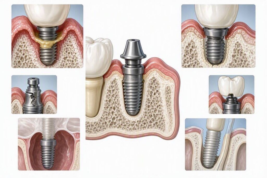
Peri-implantitis is the most significant long-term biological threat to implant survival. It is a pathological condition occurring in the tissues around osseointegrated implants. It is characterized by inflammation of the peri-implant mucosa and progressive loss of supporting bone.
The disease process begins with peri-implant mucositis, a reversible inflammatory condition confined to the soft tissue, analogous to gingivitis around natural teeth. The mucosa bleeds on gentle probing, is red and swollen, but no bone loss has occurred. If the bacterial biofilm is not effectively disrupted through professional debridement and improved patient home care, the inflammation extends apically. The host immune response destroys the bone supporting the implant.
As the bone resorbs, a deep peri-implant pocket forms. The probe drops 6, 8, or 10 millimeters or more. Pus may be expressed from the pocket. The patient notices bleeding, a bad taste, and sometimes dull pain. Radiographically, a crater-shaped bone defect appears, often saucer-shaped around the implant neck. Unlike periodontitis around a tooth, peri-implantitis can progress more rapidly because the implant lacks a periodontal ligament with a protective vascular network.
Risk factors include a history of periodontitis, poor oral hygiene, smoking, diabetes, a lack of regular professional maintenance, poorly contoured restorations that trap plaque, and residual cement from a cemented crown. Treatment ranges from nonsurgical debridement with specialized instruments to surgical access with implant surface decontamination and bone grafting. Advanced, untreated peri-implantitis leads to implant mobility and loss.
Soft Tissue Dehiscence and Mucosal Recession
The quality and quantity of the attached keratinized mucosa around an implant influence its long-term health. If the implant is placed in an area of thin, non-keratinized, mobile alveolar mucosa, the tissue may be more susceptible to inflammation and recession.
Mucosal recession exposes the gray, metallic implant collar or abutment. This is an aesthetic disaster in the anterior aesthetic zone. The patient sees a dark gray line at the gum margin. Recession also creates a plaque-retentive environment that is difficult to clean. Causes include a thin tissue biotype, a buccally positioned implant with insufficient bone on the facial aspect, a poorly contoured restoration, or traumatic toothbrushing.
Prevention involves careful three-dimensional implant placement within the bone envelope and, when indicated, soft tissue grafting to increase the width of keratinized tissue. Treatment of established recession may require a connective tissue graft surgery to cover the exposed implant surface, but the results are less predictable than root coverage on natural teeth.
The Mechanical and Technical Complications
Implants are man-made devices subjected to millions of loading cycles over years of function. Mechanical components can and do fail.
Screw Loosening and Fracture
The implant system comprises multiple components connected by tiny screws. The abutment screw secures the abutment to the implant body. The prosthetic screw secures the crown to the abutment. These screws are torqued to a precise specification, typically 15 to 35 Newton-centimeters.
Abutment screw loosening is the most common mechanical complication. The patient notices a slight mobility or a clicking sensation in the crown. The crown may feel “soft” when biting. If detected early, the screw is simply retorqued to the correct specification. If ignored, the loose screw fatigues and fractures. A fractured abutment screw is a more complex problem. The remnant of the screw remains deep inside the implant body.
Retrieving a fractured screw requires specialized retrieval kits. The surgeon or restorative dentist uses a fine drill under magnification to create a purchase point in the broken screw, then engages a reverse-threaded removal tool to back the fragment out. If retrieval is impossible, the implant may need to be removed and replaced. Screw fracture is largely preventable through proper torque protocols, passive fit of the restoration, and management of occlusal forces.
Implant Fixture Fracture
The implant body itself can fracture, although this is rare with modern titanium and zirconia implants. Fracture is almost always associated with biomechanical overload. A patient with heavy bite forces, parafunctional habits like grinding or clenching, or an implant that was placed in an off-axis loading position is at risk.
An implant fracture is a catastrophic event. The implant separates, often at the level of the first thread. The apical fragment remains osseointegrated in the bone, while the coronal fragment is mobile. The mobile fragment is removed. The embedded fragment is managed either by trephination removal or by leaving it as a sleeping implant, bypassed by a new implant. Prevention focuses on proper implant size selection, avoiding cantilever forces, and protecting the restoration with an occlusal guard.
Porcelain or Framework Fracture
The visible crown placed on the implant is subject to the same failure modes as any dental crown. Porcelain can chip or fracture from the underlying metal or zirconia framework. A minor chip can sometimes be polished or repaired with composite resin. A major fracture requires crown replacement.
Full-zirconia implant crowns have largely eliminated the porcelain chipping problem, but they are not indestructible. A severe occlusal overload can fracture a monolithic zirconia crown. The implant itself usually survives, but the crown is lost.
The Aesthetic Complications
An implant that is functionally successful—osseointegrated and pain-free—can be an aesthetic failure. In the visible smile zone, an unnatural appearance is a significant patient complaint.
The Gray Gingival Show-Through
Titanium is dark gray. If the gum tissue over the implant is thin, the underlying metal can show through, giving the gums a gray, bluish, or shadowed appearance. This is particularly noticeable if the patient has a high smile line that reveals the gum margin. Solutions include using a zirconia implant in the aesthetic zone, performing a connective tissue graft to thicken the overlying tissue, or selecting a titanium implant with a tissue-colored anodized collar.
Poor Crown Contour and Emergence Profile
A natural tooth emerges from the gum with a specific contour. An implant crown that is poorly shaped—too bulky, too narrow, or with a flat, unnatural emergence profile—looks artificial. The papilla, the triangular piece of gum tissue between teeth, may be absent, creating a “black triangle” that is aesthetically and phonetically displeasing.
Proper three-dimensional implant placement and a custom, labially contoured provisional restoration that shapes the soft tissue during the healing phase are the keys to a natural emergence profile. Correcting an established poor contour often requires removing the crown, placing a new provisional to re-sculpt the tissue, and fabricating a new custom crown.
Mid-Facial Recession
As described under biological complications, recession that exposes the implant margin is a common aesthetic failure. Once the implant collar is visible, complete correction is difficult. Soft tissue grafting to cover the exposure is a surgical attempt at camouflaging the defect, not a guaranteed solution.
The Surgical and Postoperative Complications
Complications arising from the implant surgery itself are rare when performed by a skilled, experienced surgeon, but they are serious when they occur.
Nerve Injury
The inferior alveolar nerve (IAN), which provides sensation to the lower lip and chin, runs within the mandibular canal in the lower jaw. The mental nerve branches from the IAN and exits through the mental foramen near the premolars. If an implant in the posterior mandible is placed too deep, it can encroach upon, compress, or transect the IAN.
The result is paresthesia (altered sensation, numbness, tingling) or, in severe cases, anesthesia (complete loss of sensation) of the lower lip and chin on the affected side. This can be a temporary neuropraxia from compression or a permanent nerve injury from transection.
Prevention is achieved through meticulous CBCT planning. The surgeon identifies the exact position of the canal and places the implant with a safety zone of at least 2 millimeters above the canal. If a post-operative CBCT shows implant impingement into the canal, immediate removal of the implant gives the best chance for nerve recovery. Delayed removal may result in permanent numbness.
Maxillary Sinus Perforation and Sinusitis
The maxillary sinus is an air-filled space located above the upper posterior teeth. Placing an implant in the posterior maxilla often requires a sinus lift procedure if the bone height is insufficient. Even without a sinus lift, an implant can accidentally perforate the sinus floor.
A minor perforation of the Schneiderian membrane during a sinus lift is a common intraoperative event and is managed immediately with a collagen membrane patch. It rarely causes long-term issues. However, a significant perforation or an implant that displaces into the sinus cavity is a serious complication. The displaced implant acts as a foreign body, causing chronic sinusitis, infection, and obstruction. The patient experiences unilateral nasal congestion, foul-smelling discharge, and facial pressure.
Retrieval of an implant from the sinus requires an endoscopic sinus surgery by an oral surgeon or an ear, nose, and throat specialist. Prevention is achieved through careful planning and, when necessary, a staged sinus lift procedure before implant placement.
Postoperative Infection
Acute infection at the surgical site is uncommon due to the jaw’s rich blood supply. When it occurs, it presents as increasing pain, swelling, purulent drainage, and sometimes fever several days after the surgery. A periapical abscess can form at the apex of a recently placed implant.
Management includes antibiotics, drainage, and sometimes removal of the implant if the infection cannot be controlled. Systemic factors like diabetes and smoking elevate infection risk. A sterile surgical technique and a preoperative chlorhexidine rinse are standard preventive measures.
Excessive Bleeding and Hematoma
Postoperative bleeding is typically self-limited and managed with gauze pressure. However, bleeding can be more significant if the implant osteotomy severs a branch of the facial or lingual artery, particularly in the floor of the mouth for mandibular anterior implants. A sublingual hematoma can expand and compromise the airway, constituting a surgical emergency. This is an extremely rare but life-threatening complication. The surgeon must be aware of the lingual anatomy and plan the implant angulation accordingly.
The Systemic and Patient-Related Complications
The patient’s systemic health and behaviors directly influence implant outcomes.
Implant Failure in Patients with Uncontrolled Diabetes
Chronic hyperglycemia impairs wound healing, reduces neutrophil function, and increases susceptibility to infection. Patients with poorly controlled diabetes, indicated by a hemoglobin A1c above 7% or 8%, have a significantly higher risk of early implant failure and peri-implantitis. The implant surgeon will require a recent A1c result and may coordinate care with the patient’s endocrinologist before proceeding.
Bisphosphonate-Related Osteonecrosis of the Jaw (BRONJ)
Patients taking intravenous bisphosphonates (such as zoledronic acid) for bone metastases or multiple myeloma, and, to a lesser extent, patients on long-term oral bisphosphonates for osteoporosis, are at risk for osteonecrosis of the jaw. A dental extraction or implant surgery can trigger a non-healing, exposed area of necrotic bone. This is a devastating and difficult-to-manage complication.
The implant surgeon must take a detailed medication history. Patients on IV bisphosphonates are generally not candidates for elective implant surgery. Patients on oral bisphosphonates require a discussion with their physician about the risks, a drug holiday may be considered, and the risk of BRONJ must be clearly communicated and documented in the consent.
Allergic Reaction to Implant Materials
Titanium allergy is exceedingly rare but exists. Cases of allergic contact dermatitis, implant failure, and localized eczematous reactions attributed to titanium hypersensitivity are reported in the literature. Zirconia implants are a metal-free alternative for the very small population of patients with demonstrated titanium allergy or those who request metal-free treatment for personal reasons.
Conclusion
Dental implants can go wrong in biological, mechanical, aesthetic, and surgical domains, with peri-implantitis and screw loosening being the most frequent long-term complications, while nerve injury and sinus perforation represent rare but serious surgical risks. The majority of complications are preventable through meticulous three-dimensional CBCT planning, a sterile surgical technique, proper prosthetic design that manages occlusal forces, and a lifelong commitment to professional maintenance and home care. A patient who understands these risks, selects an experienced surgeon, and complies with the maintenance protocol has an excellent probability of a successful, decades-long implant service life.
Frequently Asked Questions
How common is peri-implantitis?
Studies report varying prevalence rates, but systematic reviews suggest that peri-implantitis affects approximately 10% to 20% of implants and 18% to 22% of patients over a five- to ten-year period. The wide range reflects different diagnostic criteria. The key message is that peri-implantitis is common enough that every implant patient must be enrolled in a regular professional maintenance program and practice meticulous home care.
Can a body reject a dental implant?
True immunologic rejection, as seen with organ transplants, does not occur with titanium or zirconia implants. The body does not produce antibodies against the implant. What is perceived as “rejection” is almost always a failure of osseointegration due to infection, surgical technique, or overload. Titanium allergy is a rare, localized hypersensitivity, not a systemic rejection.
Is it safe to get an implant if I smoke?
Smoking significantly impairs healing and increases the risk of both early implant failure and peri-implantitis. Smokers have a failure rate approximately two to three times higher than non-smokers. Many implant surgeons will advise a smoking cessation period before and after surgery. Some decline to place implants in heavy smokers due to the unacceptably high risk. Quitting smoking is the single most impactful patient-modifiable risk reduction strategy.
What are the signs that my dental implant is failing?
The warning signs include bleeding when brushing around the implant, swollen or red gums around the implant, a bad taste or pus discharge, increasing mobility of the crown or the implant, pain or discomfort when biting, and a visible gray line at the gum margin. If you notice any of these, contact your implant dentist immediately. Early intervention for peri-implant mucositis can reverse the condition before bone loss occurs.
Additional Resource
The International Team for Implantology (ITI) provides a free, peer-reviewed patient education section at iti.org. The ITI is a global academic network of implant dentistry experts. Their patient resources explain implant treatment, complications, and maintenance in accessible language grounded in the latest scientific evidence, making it an authoritative supplement to your surgeon’s specific guidance.