What Holds Dental Implants In Place?
Dental implants stay in place through a remarkable biological phenomenon called osseointegration. Unlike dentures that rely on suction or adhesives, and unlike bridges that depend on adjacent teeth for support, dental implants are anchored directly by living bone. This article explains the science of implant retention, the factors that influence successful osseointegration, the mechanical connections that secure the restoration to the implant, and what patients can do to maintain implant stability for decades.
The Foundation: Osseointegration
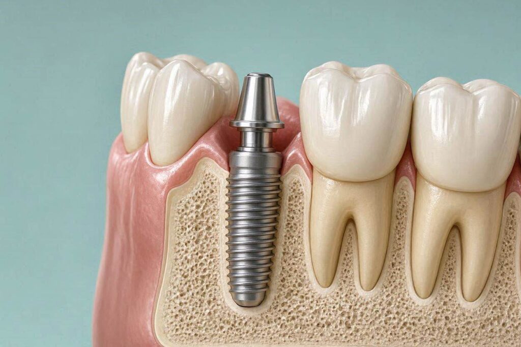
Osseointegration is the direct structural and functional connection between living bone and the surface of a load-bearing implant. This biological process is what fundamentally holds a dental implant in place.
The Discovery of Osseointegration
Swedish orthopedic surgeon Per-Ingvar Brånemark discovered osseointegration serendipitously in the 1950s while studying blood flow in rabbit bone. He placed titanium optical chambers into the bone of experimental animals. When he attempted to remove these chambers at the conclusion of his studies, he found that the bone had fused so tightly to the titanium that the chambers could not be removed without fracturing the surrounding bone.
Brånemark recognized the clinical potential of this phenomenon. He shifted his research focus and eventually developed the first titanium dental implants. The first human patient received Brånemark implants in 1965. Those implants remained functional for over 40 years until the patient’s death, a testament to the durability of osseointegration.
How Osseointegration Works at the Cellular Level
When a titanium implant is placed into prepared bone, a sequence of biological events unfolds. Within seconds of placement, blood proteins adsorb onto the implant surface, forming a conditioning film. Platelets adhere and release growth factors that attract bone-forming cells.
Osteoblasts, the cells responsible for bone formation, migrate to the implant surface. They begin laying down bone matrix directly onto the titanium oxide layer. Over weeks and months, this woven bone matures and remodels into lamellar bone, organized and strong. The bone grows into the micro-roughness of the implant surface, creating mechanical interlocking at the microscopic level.
The resulting bone-implant interface is not a fibrous encapsulation, which would indicate failure, but a direct bone-to-implant contact. This structural continuity means forces applied to the implant during chewing are transmitted directly to the bone, stimulating bone maintenance in the same way natural tooth roots do.
The Importance of the Implant Surface
The surface characteristics of the implant significantly influence osseointegration. Modern implants undergo surface treatments to optimize bone response.
Machined titanium surfaces, used in early implants, were relatively smooth. Healing times were longer, and success rates, while good, were lower than current standards. Contemporary implants feature moderately rough surfaces created through sandblasting, acid-etching, or anodization. These roughened surfaces provide greater surface area for bone contact and promote more rapid and robust osseointegration.
Some implant surfaces are chemically modified to be hydrophilic, attracting water and blood, which accelerates the early healing response. Others incorporate calcium phosphate or other bioactive coatings that stimulate bone formation. These surface technologies have reduced healing times from the original six months to as little as three to four weeks in favorable conditions.
The Role of Primary Stability
Osseointegration takes time. During the healing period, the implant must be held motionless by mechanical means. This initial mechanical retention is called primary stability.
How Primary Stability is Achieved
The surgeon prepares an osteotomy, a precisely shaped hole in the bone, slightly smaller in diameter than the implant. When the threaded implant is screwed into this undersized preparation, it compresses the surrounding bone. The threads engage the bone walls, creating a tight mechanical fit.
The surgeon measures insertion torque during placement. Values between 30 and 50 Newton-centimeters indicate good primary stability. Higher insertion torque values suggest the implant is firmly anchored. Inadequate primary stability, with low insertion torque, predicts higher failure risk because micromotion during healing can prevent osseointegration.
The Transition from Primary to Secondary Stability
In the days and weeks following placement, primary stability actually decreases somewhat as the compressed bone remodels and the initial mechanical interlock relaxes. Simultaneously, secondary stability, the biological fixation from new bone formation, increases. There is a critical period, typically around two to four weeks post-placement, when total stability dips as primary stability declines faster than secondary stability develops. Avoiding loading during this period protects the implant.
By three to six months, osseointegration is mature, and the implant is anchored by living bone, not merely by mechanical friction. At this point, the implant can bear functional loads.
The Mechanical Components Above the Bone
While osseointegration holds the implant fixture in the bone, additional mechanical connections secure the restoration to the implant.
The Implant-Abutment Connection
The abutment connects to the implant fixture through one of several connection designs. The most common is the internal conical connection, where the abutment has a tapered portion that fits into a matching taper within the implant. This connection provides a friction fit that seals the interface against bacterial leakage.
External hex connections, an older design, locate the abutment on an external hexagon on top of the implant. These connections are simpler but may be more susceptible to screw loosening. Internal hex, octagon, and tri-lobe connections provide anti-rotational features combined with varying degrees of connection stability.
The abutment is secured to the implant with an abutment screw. This screw is tightened to a precise torque, typically 25 to 35 Newton-centimeters, using a torque wrench. Proper torque is critical. Under-tightening leads to screw loosening. Over-tightening can strip threads or fracture the screw.
The Crown-Abutment Connection
The crown attaches to the abutment either through cement retention or screw retention. In cement-retained restorations, the crown is bonded to the abutment with dental cement. The cement fills the space between the internal surface of the crown and the external surface of the abutment, creating a mechanical bond.
In screw-retained restorations, the crown has an access channel through which a screw passes directly into the abutment or implant. The screw secures the crown in place. The access channel is then filled with composite material to seal it and restore the occlusal surface.
The Role of Splinting
When multiple implants support a single prosthesis, as in a full-arch restoration, the prosthesis splints the implants together. This splinting distributes forces across all the supporting implants. If one implant were to develop problems, the splinted prosthesis shares the load among the remaining implants. The metal framework or zirconia structure of the prosthesis provides rigidity.
Factors That Maintain or Threaten Implant Stability
Long-term implant retention depends on patient factors and habits.
Factors That Maintain Stability
Healthy peri-implant tissues maintain the soft tissue seal that protects underlying bone. Meticulous oral hygiene, including daily brushing, interdental cleaning, and antimicrobial rinsing as recommended, prevents peri-implant inflammation. Regular professional maintenance visits allow early detection of problems before they compromise stability.
A balanced occlusion distributes forces appropriately. The dentist adjusts the bite to ensure the implant crown does not bear excessive or eccentric forces. Night guards protect implants from bruxism forces during sleep.
Adequate bone volume and quality at the time of placement provide the foundation for long-term stability. Bone grafting, when needed, creates a better foundation.
Factors That Threaten Stability
Peri-implantitis, the inflammatory destruction of bone around an implant, is the leading cause of late implant instability. Bacterial plaque, combined with host inflammatory response, progressively destroys the bone that holds the implant. Untreated, this leads to mobility and implant loss.
Excessive occlusal forces, from a high restoration, parafunctional habits, or an unbalanced bite, can exceed the bone’s capacity to adapt. Bone loss around the implant may result, leading to instability.
Poor bone quality, such as very soft or sparse bone, may not provide adequate support for osseointegration. Medical conditions including uncontrolled diabetes, immunocompromise, and certain medications can impair bone healing and maintenance.
Smoking and nicotine use impair blood flow, immune response, and bone metabolism. Smokers experience significantly higher rates of implant failure due to compromised osseointegration and increased peri-implantitis risk.
What Holds Implant Overdentures in Place
Implant overdentures combine biological fixation with mechanical retention.
The Attachment System
Overdentures use attachment systems that connect the removable denture to the implant abutments. The abutments are screwed into the implants and have patrix components, such as locator studs or ball attachments, that protrude through the gum.
The denture base contains corresponding matrix components, such as nylon inserts or O-rings, that snap onto the patrix abutments. This mechanical retention holds the denture in place during function. The patient overcomes the retention force to remove the denture for cleaning.
The Dual Retention Mechanism
The overdenture is held in place by both the mechanical attachment to the implants and the tissue support of the denture base on the residual ridge. The implants provide retention and stability against vertical and horizontal displacement. The ridge provides support against vertical forces. This dual mechanism significantly improves function compared to conventional dentures while still allowing removal.
What Happens When an Implant Loses Its Hold
Implant mobility is the definitive sign that osseointegration has failed.
Early Failure
Early failure occurs before or at the time of restoration. The implant never achieved osseointegration or lost it during the healing period. The implant is mobile, often without significant pain. It must be removed. The site is allowed to heal, and replacement is planned after addressing the factors that led to failure.
Late Failure
Late failure occurs after a period of successful function. Peri-implantitis, occlusal overload, or implant fracture can cause late failure. The implant becomes mobile as supporting bone is lost. In advanced cases, the implant may be easily removed, reflecting the complete destruction of the bone-implant interface.
The Difference Between Implant Mobility and Crown Mobility
Not all perceived mobility means the implant has failed. A loose abutment screw produces mobility of the crown while the implant remains solidly integrated. A loose crown on its abutment also produces crown mobility. The dentist distinguishes between these scenarios. Implant mobility is diagnosed by directly assessing the implant fixture. Crown or abutment mobility is diagnosed by manipulating the restoration components.
Conclusion
Dental implants are held in place primarily by osseointegration, the direct biological bonding of living bone to the implant surface. This process, discovered by Brånemark and refined through decades of research, creates a structural connection that transmits functional forces to the bone, preserving it. Primary mechanical stability at the time of placement provides the immobility necessary for osseointegration to occur. The abutment screw and crown cement or screw provide the mechanical connections above the bone. Long-term stability depends on maintaining healthy peri-implant tissues, balanced occlusal forces, and good systemic health. When osseointegration fails, the implant becomes mobile and requires removal.
Frequently Asked Questions
Can an implant come loose after years of being stable?
Yes. Late implant failure can occur due to peri-implantitis, occlusal overload, or implant fracture. Regular maintenance visits detect problems before they progress to mobility.
How strong is the bond between bone and implant?
The bone-implant interface is remarkably strong. Removal of a well-integrated implant requires significant force or surgical trephination. The bond withstands the substantial forces of chewing for decades.
What is the difference between a loose implant and a loose crown?
A loose crown or abutment screw produces crown mobility while the implant remains solid. A loose implant means the implant fixture itself moves within the bone. The dentist distinguishes between these using specific diagnostic tests.
Can a loose implant be tightened?
No. An implant that has lost osseointegration cannot be tightened. The biological attachment has failed. The implant requires removal. Mechanical components like abutment screws can be tightened.
Additional Resource
For scientific information on osseointegration and implant dentistry, visit the Academy of Osseointegration at www.osseo.org. The Academy provides professional and patient education on the biological basis of implant retention.