Are Veneers Safe Long Term?
The veneer consultation is a seduction. The dentist shows you a digital smile design, a preview of perfectly proportioned, brilliantly white teeth. The promise is a lifetime of confidence. The paperwork you sign mentions “irreversible enamel removal” and “future replacement,” but those words float past, drowned out by the allure of the before-and-after gallery. Then, 10, 15, or 20 years later, the bill for that seduction comes due, and the question that was glossed over in the excitement suddenly becomes urgent: “Are veneers safe long term?”
Safety in this context is not a single variable. It is a composite of structural integrity (will the tooth underneath rot?), periodontal health (will the gums recede and expose a dark margin?), biological compatibility (will the pulp die?), functional stability (will the bite collapse?), and aesthetic durability (will the smile still look natural in two decades?). The honest answer, grounded in the longitudinal literature and clinical experience, is that well-executed porcelain veneers on properly selected patients are a safe, durable, and predictable restoration with a 15–25 year lifespan, but they are not biologically inert and they are not permanent. They are a one-way door. The tooth underneath is permanently altered, and the restoration will require maintenance, replacement, and eventual financial reinvestment for the rest of the patient’s life. This article maps the long-term biological risks, the structural failure modes, the periodontal consequences, and the honest maintenance burden that every prospective veneer patient must internalize.
The Irreversibility Principle: The Foundational Truth
Every discussion of long-term veneer safety must begin with the single most important biological fact: enamel does not regenerate. The preparation of a tooth for a porcelain veneer involves the removal of approximately 0.3 to 0.7 millimeters of enamel from the facial surface and, in most cases, the incisal edge. This enamel is gone forever. The tooth is now a reduced, vulnerable substrate that will never again exist in its natural, intact state.
This is not inherently unsafe. The veneer replaces the removed enamel with a layer of porcelain that is bonded to the tooth with resin cement, and this porcelain-resin-enamel-dentin complex is structurally sound and biologically compatible. But it is a prosthetic replacement, not a biological restoration. If the veneer debonds, fractures, or is removed for any reason, the tooth underneath cannot be left exposed. It must be immediately re-covered with a new restoration. There is no “going back to natural.” The patient who chooses veneers is making a lifelong commitment to maintaining a prosthetic covering on those teeth. The safety question, therefore, is not “Are veneers safe?” but “Is the lifetime prosthetic maintenance burden acceptable and manageable?”
The Margin: Where Safety Succeeds or Fails
The critical biological interface in a veneer is the margin—the microscopic line where the porcelain edge meets the tooth structure, usually at or just below the gumline. Long-term safety is overwhelmingly determined by the quality of this margin and the patient’s ability to keep it clean.
A perfectly executed veneer margin, placed by a skilled dentist on sound enamel, polished to a sub-micron smoothness, and bonded with a hydrophobic resin cement under absolute moisture control, creates a seamless transition that resists bacterial penetration. The margin is cleansible. A toothbrush bristle can sweep across it without catching. Bacteria cannot easily colonize the interface.
A poorly executed margin—over-contoured, placed too deeply subgingivally, left with a ledge or an open gap, or bonded in a contaminated field—creates a plaque trap. Bacteria colonize the gap. The biofilm matures. Acids demineralize the enamel at the margin. Decay begins, usually invisibly, underneath the veneer. This is “margin decay,” the most common long-term failure mode of veneers. The decay progresses silently, hidden by the porcelain, until the veneer debonds because the underlying tooth structure has rotted away, or the patient presents with sensitivity and radiographic evidence of a carious lesion under the restoration. The safety of the veneer over decades is a direct function of the margin seal. A well-sealed margin on a patient with excellent oral hygiene is biologically safe. An open margin on a patient who skips flossing is a slow-motion dental catastrophe.
Periodontal Health: The Gum Response
The relationship between the veneer margin and the gingival tissues is another long-term safety variable. Porcelain is biocompatible. It is inert, non-porous, and well-tolerated by the periodontal tissues. In fact, a highly polished porcelain surface is less plaque-retentive than natural enamel. However, the margin—the junction of porcelain and tooth—is a disruption in the smooth surface contour, and it can harbor bacteria if it is not perfectly finished.
An over-contoured veneer that bulges at the gumline creates a “plaque shelf.” The gingival tissues respond with inflammation—redness, swelling, bleeding on probing. Chronic marginal gingivitis, if left untreated, can progress to periodontitis, with attachment loss and gingival recession. The recession exposes the veneer margin and, critically, may expose the underlying discolored tooth structure or the dark line of the resin cement. This is the “dark margin” aesthetic failure that is often blamed on the veneer but is actually a periodontal problem.
The long-term periodontal safety of veneers depends on three factors: the biologic width is not violated during preparation (the margin should not extend more than 0.5 millimeters into the gingival sulcus), the emergence profile is anatomically contoured, and the patient maintains meticulous interproximal hygiene. A patient who flosses daily, uses an interdental brush, and attends regular hygiene visits can maintain healthy gingival tissues around veneers for decades. A patient who neglects hygiene will develop inflammation, recession, and margin exposure. The veneer is a passive participant in this process; the active variable is the host’s immune-inflammatory response to bacterial plaque.
Pulpal Health: The Nerve Inside the Tooth
The dental pulp—the living nerve and blood supply inside the tooth—can be injured by the veneer preparation process. The heat generated by the high-speed handpiece during enamel reduction, the vibration, and the desiccation of the dentin during bonding can all cause transient or permanent pulpal inflammation. In the vast majority of cases, this inflammation is mild, reversible, and resolves without consequence. The tooth remains vital (alive) and asymptomatic for the life of the patient.
However, a small percentage of veneered teeth—estimates range from 1% to 5% in the literature, depending on the depth of preparation and the proximity to the pulp—will undergo pulpal necrosis (death of the nerve) in the months or years following veneer placement. The tooth may become spontaneously painful, sensitive to hot or cold, or develop a periapical abscess visible on a radiograph. This is not a “failed veneer”; it is a biological consequence of the preparation trauma. The tooth requires root canal therapy, which involves accessing the pulp chamber through the back of the tooth (preserving the veneer on the facial surface) or, in some cases, through the veneer itself, which then requires replacement.
Long-term pulpal safety is optimized by conservative preparation depth—staying within enamel as much as possible, avoiding aggressive dentin exposure—and by using copious water spray during preparation to dissipate heat. Patients with pre-existing large restorations, deep decay, or a history of trauma to the teeth are at higher risk for pulpal complications and should be informed of this risk before consenting to elective veneers.
Structural Failure: Fracture, Debond, and Wear
Porcelain is a brittle material. It is strong in compression but weak in tension. A veneer that is well-supported by the underlying tooth structure and bonded with modern resin cement is remarkably resistant to fracture under normal masticatory forces. However, parafunctional habits—bruxism (grinding), clenching, nail-biting, pen-chewing, ice-chewing—subject veneers to extreme lateral and tensile forces that can cause catastrophic porcelain fracture.
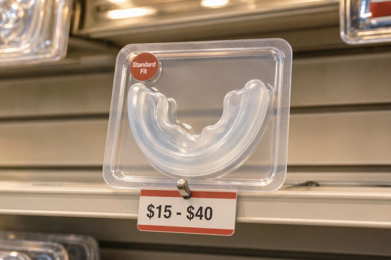
A patient with untreated nocturnal bruxism who receives maxillary porcelain veneers is at high risk of fracturing the incisal edges. The porcelain chips, cracks, or shears off, exposing the underlying tooth. The veneer must be replaced. A properly fabricated occlusal guard (night guard) worn consistently is the essential protective measure for the bruxing veneer patient. Without it, the long-term structural safety of the veneers is severely compromised.
Debonding—the veneer coming off the tooth in one piece—is less common with modern adhesive protocols but still occurs. Debonding is usually an adhesive failure at the resin-tooth interface, caused by contamination during bonding, inadequate enamel etching, or excessive force. A debonded veneer can often be re-bonded if it is intact and the tooth surface is clean. A fractured veneer cannot be repaired and must be replaced.
Occlusal wear of the porcelain against the opposing natural teeth is another consideration. Porcelain is harder than enamel and can abrade the opposing natural dentition if the occlusion is not carefully adjusted. This is a long-term functional safety issue: the veneers may be intact, but the opposing teeth may be worn down, requiring restorative intervention.
Aesthetic Longevity: The Fading Smile
Porcelain itself is color-stable. It does not stain, yellow, or darken over time. This is one of its great advantages over composite resin bonding. However, the resin cement layer that bonds the veneer to the tooth can discolor over many years, particularly if the margin is exposed or if the cement is a conventional light-cure composite that is susceptible to water sorption and color change. The veneer may appear to “yellow” when it is actually the cement layer darkening beneath the translucent porcelain.
The soft tissue aesthetics also change over decades. Gingival recession exposes the veneer margin and the underlying tooth structure, creating a visible line. The lip position changes with aging—the upper lip droops, showing less of the maxillary teeth at rest. The smile that was perfectly framed by the lips at age 30 may look different at age 60. The veneers are static; the face is dynamic. This is not a “failure” of the veneers, but it is a long-term aesthetic consideration that may prompt replacement for purely cosmetic reasons.
The Replacement Cycle: 15–20 Years and Beyond
The question “Are veneers safe long term?” inevitably leads to “How long do they last?” The literature reports survival rates of 90–95% at 10 years and 80–90% at 20 years for well-executed porcelain veneers. This means that 1 in 10 to 1 in 5 veneers will require replacement within two decades. The reasons for replacement are margin decay, porcelain fracture, aesthetic dissatisfaction (color, shape, recession), debonding, and pulpal complications.
Replacement is more complex and more biologically costly than the initial placement. The old veneer must be removed, which usually requires some degree of further tooth reduction as the residual cement and a thin layer of enamel are ground away. The tooth is now slightly smaller. The new veneer is placed on a more compromised substrate. Each replacement cycle incrementally reduces the remaining tooth structure, bringing the preparation closer to the pulp and increasing the risk of pulpal necrosis and the eventual need for a full crown or root canal therapy. After two or three replacement cycles over a lifetime, a tooth that began with a conservative veneer preparation may end up needing a full-coverage crown. This is the long-term biological trajectory. It is not a failure of the initial treatment; it is the inevitable consequence of the irreversible nature of enamel removal.
Comparative Table: 10-Year vs. 20-Year Veneer Outcomes
| Parameter | 10-Year Outcome | 20-Year Outcome |
|---|---|---|
| Survival Rate | 90–95% of veneers still in situ. | 80–90% still in situ; some replaced. |
| Margin Integrity | Generally intact; minor staining possible. | Margin staining, decay, or gap formation in a minority. |
| Gingival Health | Healthy if hygiene maintained; minor recession possible. | Recession visible; margin may be exposed. |
| Pulpal Vitality | Over 95% of teeth remain vital. | 90–95% vital; small percentage require root canal. |
| Aesthetic Satisfaction | High; porcelain color stable. | Moderate to high; cement discoloration, soft tissue aging. |
| Opposing Tooth Wear | Minimal if occlusion adjusted. | Accelerated wear if bruxism or malocclusion present. |
Important Note: The “No-Prep” Long-Term Unknown
Thin, no-prep or minimal-prep veneers (0.2–0.3 millimeters) have been marketed as a safer, less invasive alternative with a reversible profile. The long-term data on these restorations is less robust than for traditional veneers. The very thin porcelain is more prone to fracture, and the bond to un-prepared enamel, while excellent, is under higher stress because the restoration is thin. Additionally, no-prep veneers add thickness to the tooth, which can create an over-contoured emergence profile that traps plaque and irritates the gingiva. The long-term periodontal safety of no-prep veneers is a subject of ongoing study, and the “reversible” claim should be viewed skeptically—even if no enamel was removed, the bonding process alters the enamel surface, and removal of the veneer still requires grinding off the porcelain and cement, which inevitably removes some enamel. True reversibility is a marketing claim, not a biological reality.
Conclusion
Porcelain veneers are a safe, biologically compatible long-term restoration with 15–25 year survival rates exceeding 80%, provided the margins are sealed perfectly on sound enamel, the patient maintains meticulous oral hygiene and periodontal health, and parafunctional habits like bruxism are controlled with an occlusal guard. The irreversible removal of enamel commits the patient to a lifetime of prosthetic maintenance, with each successive replacement cycle consuming incremental tooth structure and increasing the risk of pulpal necrosis and eventual full-coverage restoration. The long-term safety question is not “Will these last forever?” but “Am I prepared for the lifelong financial and biological maintenance that these beautiful shells demand?”
FAQ
Q: Can veneers cause tooth decay underneath?
A: Veneers themselves do not cause decay. Decay occurs at the margin—the junction between the porcelain and the tooth—when bacteria colonize a gap, an open margin, or a rough cement line. If the veneer margin is perfectly sealed and the patient maintains excellent oral hygiene, the risk of decay is very low. If the margin is defective or hygiene is poor, decay can progress silently under the veneer, often undetected until it is extensive. Regular dental exams with bitewing radiographs are essential for detecting margin decay early.
Q: Do veneers make your teeth more sensitive long term?
A: Most post-veneer sensitivity is transient and resolves within days to weeks after the preparation. Long-term, a properly bonded veneer should not cause sensitivity; the porcelain and cement seal the dentin tubules. If a veneered tooth develops persistent sensitivity years later, it is a red flag for margin leakage, recurrent decay, cement washout, or a crack in the tooth or porcelain. It requires immediate evaluation. Do not ignore a tooth that suddenly becomes sensitive years after veneers were placed.
Q: What happens to my teeth if I decide I don’t want veneers anymore after 15 years?
A: You cannot simply remove veneers and leave the teeth bare. The enamel has been removed, and the exposed tooth surface is dentin, which is softer, more yellow, more sensitive, and more decay-prone than enamel. Your only options are to replace the veneers with new veneers, place full-coverage crowns if the tooth structure is insufficient for veneers, or, in a worst-case scenario with extensive decay, extract the teeth and consider implants or a bridge. There is no “un-do” button. This is why the initial decision to proceed with veneers must be made with full understanding of the permanence.
Additional Resource
For longitudinal clinical studies on porcelain veneer survival rates and biological complications, the Journal of Esthetic and Restorative Dentistry and the American Academy of Cosmetic Dentistry (www.aacd.com) provide peer-reviewed data and patient education resources.