Can Dental Implant Detach From Bone?
The defining characteristic of a successful dental implant is osseointegration: a direct, structural, and functional connection between living bone and the surface of the titanium or zirconia implant. This biological bond is the foundation upon which the entire implant restoration depends. The question of whether this bond can fail—whether the implant can detach from the bone—is a question about the fundamental durability of the implant-bone interface. The answer is yes, an implant can detach from the bone. This detachment is the definition of implant failure, and it can occur in distinct clinical contexts at different times in the implant’s lifespan. This guide provides a comprehensive, biologically grounded explanation of how and why implant-bone detachment occurs, the difference between early and late failure, the clinical signs, and the management and prevention strategies.
The Biology of Osseointegration and Its Failure
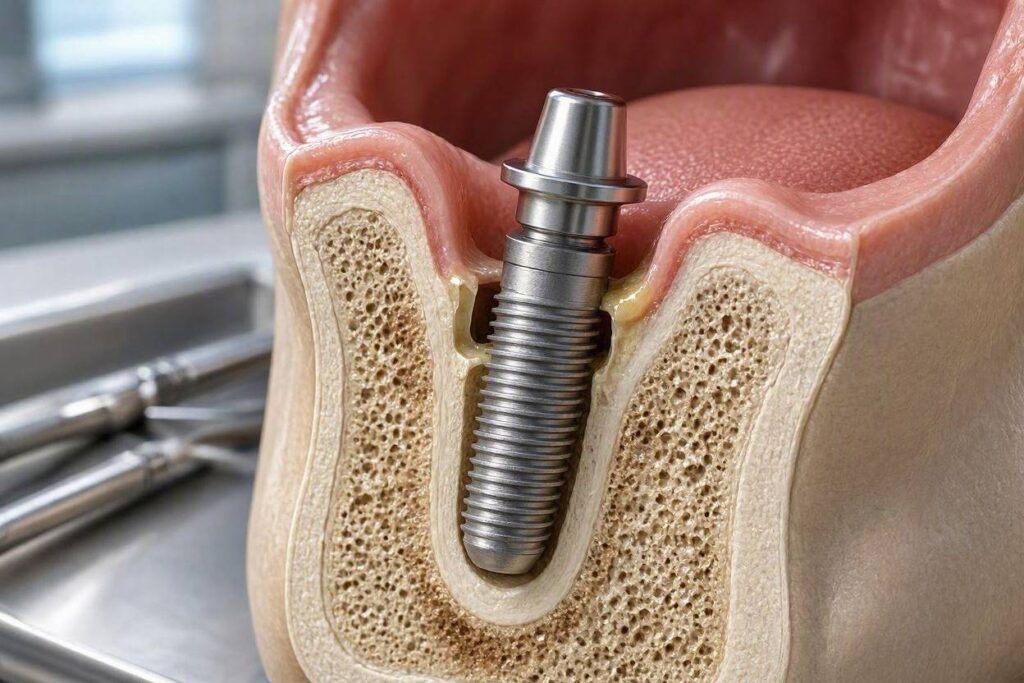
To understand detachment, one must first understand the normal process of bone attachment. When an implant is placed into a precisely prepared osteotomy in the jawbone, blood fills the space between the implant threads and the bony walls. A blood clot forms, containing platelets, growth factors, and mesenchymal stem cells. Over the initial days and weeks, this clot is replaced by granulation tissue, then by woven bone, and finally by mature, lamellar bone.
The bone does not simply grow alongside the implant. At the microscopic level, bone-forming cells (osteoblasts) deposit bone matrix directly onto the titanium oxide layer on the implant surface. The bone and the implant become interlocked at the micro- and nano-scale. This is osseointegration. The implant is not glued or cemented to the bone; it is biologically bonded. Removing an osseointegrated implant requires cutting the bone around it with a trephine bur or applying significant reverse torque to fracture the bone-implant interface.
Detachment occurs when this biological bond fails. The bone resorbs away from the implant surface, and the space is filled with inflammatory granulation tissue or fibrous connective tissue. The implant loses its anchorage and becomes mobile.
Early Detachment: Failure of Primary Osseointegration
Early detachment occurs within the first weeks to months after implant placement, before the implant has been restored with a crown. The implant is placed, and instead of bone forming on its surface, a fibrous tissue capsule forms. The implant fails to integrate.
Causes of Early Detachment
- Surgical Trauma: Overheating the bone during the drilling of the osteotomy is a primary cause. If the bone temperature exceeds 47 degrees Celsius (approximately 117 degrees Fahrenheit) for more than one minute, the bone cells (osteocytes) die. This is called thermal necrosis. The dead bone is resorbed by the body, and the implant is surrounded by non-viable tissue, not living bone. Copious irrigation with sterile saline during drilling prevents thermal necrosis.
- Implant Contamination: If the implant surface is contaminated with bacteria, surgical glove powder, or organic debris before insertion, the bone cells will not migrate to the surface. Strict sterile surgical technique is mandatory.
- Inadequate Primary Stability: If the implant does not achieve sufficient mechanical stability at the time of placement—because the bone is too soft, the osteotomy was over-prepared, or the implant design is not appropriate for the bone quality—micromotion occurs. Even microscopic movement of the implant during the healing phase prevents the bone from forming a stable attachment. The bone cells require a stable scaffold.
- Infection: Bacterial contamination of the surgical site can lead to an acute or subacute infection that destroys the forming bone-implant interface.
- Systemic Patient Factors: Uncontrolled diabetes, heavy smoking, immunosuppression, and medications that impair bone healing (such as bisphosphonates) compromise the body’s ability to form bone on the implant surface.
- Premature Loading: If the patient chews on the implant or the provisional restoration transmits forces to the implant before osseointegration is complete, the micromotion will cause fibrous encapsulation instead of bone integration.
Clinical Signs of Early Detachment
The implant is mobile when tested by the surgeon. There may be a radiolucent (dark) line surrounding the implant on an X-ray. The patient may report a dull ache or a sensation of looseness. There may be signs of infection, such as swelling, redness, or pus. The implant is non-restorable. It must be removed.
Late Detachment: Loss of Osseointegration After Function
A successfully osseointegrated implant can detach from the bone years after it has been functioning under a crown, bridge, or denture. This is late failure, and the most common cause is peri-implantitis.
Peri-Implantitis-Induced Bone Loss
Peri-implantitis is a bacterial infectious disease. Plaque biofilm accumulates on the implant surface, the abutment, and the crown margins. The host immune system responds with inflammation. Pro-inflammatory cytokines stimulate osteoclasts (bone-resorbing cells) to break down the bone surrounding the implant. The bone is progressively resorbed, starting at the crestal bone level and moving apically.
As the bone loss progresses, the implant loses its supporting bone. In the early and moderate stages, the implant remains rigid and immobile because the remaining bone in the apical portion still provides anchorage. The disease is detected by probing, bleeding, and radiographic bone loss, not by mobility.
When the bone loss reaches a critical level—when the remaining bone can no longer support the implant against occlusal forces—the implant becomes mobile. The bone-implant interface has been destroyed by the inflammatory process. The implant has effectively detached from the bone. At this stage, the implant is terminally failing and must be removed.
Biomechanical Overload and Microfractures
Excessive occlusal forces, particularly lateral (off-axis) forces, can cause microfractures at the bone-implant interface. Over time, these microfractures accumulate, and the bone resorbs as a stress-shielding response. The implant remains rigid for a period, but the progressive bone loss can eventually lead to mobility and detachment. This mechanism is often combined with peri-implantitis, as the bone loss from overload creates a niche for bacterial colonization.
Implant Fracture
A fractured implant is not biologically detached, but it is mechanically separated. The coronal fragment is mobile or has come out, while the apical fragment remains osseointegrated. This is a material failure, not a biological detachment, but the functional result is the same: the implant restoration is lost.
The Clinical Examination for Detachment
The diagnosis of implant detachment is made through a combination of clinical examination and radiographic imaging.
Mobility Testing
The surgeon uses two instrument handles to apply lateral pressure to the implant. An osseointegrated implant has zero mobility. Any perceptible movement indicates that the bone-implant bond has failed. Mobility is the definitive sign of detachment. There is no radiographic substitute for the clinical test of mobility.
Radiographic Evaluation
A periapical X-ray or a CBCT scan reveals a radiolucent line around the implant, indicating a gap between the implant surface and the bone. In early failure, the line may surround the entire implant. In late peri-implantitis, a crater-shaped bone defect is seen, beginning at the crestal bone and extending apically. The implant may still be rigid if the apical bone is intact. The radiograph alone, without mobility testing, cannot definitively diagnose detachment in all cases.
Management of a Detached Implant
A mobile implant is non-salvageable. There is no treatment that can re-establish osseointegration on a mobile implant. The implant must be removed.
Removal Procedure
The removal technique is described in detail in the dedicated guide “How Are Failed Dental Implants Removed?” A mobile implant with extensive bone loss and fibrous encapsulation can often be simply unscrewed or gently elevated. A partially integrated, non-mobile but failing implant requires trephination or piezosurgery to cut the residual bone-implant interface.
Site Management and Future Implant Placement
After removal, the residual bony defect is thoroughly debrided of all granulation tissue. If the goal is to replace the implant, a bone graft is placed to regenerate the lost bone volume. The site heals for three to six months. A new implant is placed into the healed, grafted bone. The success rate for re-implantation in a properly managed site is high.
Prevention of Implant Detachment
Preventing detachment focuses on the modifiable risk factors for failure.
- Preoperative Planning: A CBCT scan ensures that the implant is placed into adequate bone volume and quality. A surgical guide ensures precise positioning.
- Sterile Surgical Technique: Strict aseptic protocol prevents bacterial contamination.
- Atraumatic Surgery: Copious irrigation, sharp drills, and controlled drilling speed prevent thermal necrosis.
- Achieving Primary Stability: The surgeon confirms adequate insertion torque and implant stability at the time of placement.
- Patient Risk Factor Modification: Smoking cessation, glycemic control for diabetic patients, and management of parafunctional habits (night guard for bruxism) are essential.
- Meticulous Oral Hygiene and Professional Maintenance: The patient must perform the daily cleaning protocol described in the dedicated cleaning guide. Professional maintenance every three to six months with non-metal instruments detects early peri-implantitis before it progresses to bone loss and detachment.
Conclusion
A dental implant can detach from bone, and this detachment is the definition of implant failure. Early detachment, within weeks to months of placement, results from a failure of primary osseointegration due to surgical trauma, contamination, infection, inadequate stability, or systemic patient factors. Late detachment, occurring years after function, is caused by progressive bone loss from peri-implantitis or biomechanical overload, eventually destroying the bone-implant interface to the point of mobility. A mobile implant is non-salvageable and must be removed, after which the site can be grafted and a new implant placed after a healing period.
Frequently Asked Questions
Can an implant detach from the bone and then reattach on its own?
No. There is no spontaneous re-osseointegration of a mobile implant. Once the bone-implant bond is broken and the implant is mobile, the space fills with inflammatory tissue, and reattachment is biologically impossible. The implant must be removed.
Is implant detachment painful?
Early detachment from failed osseointegration is often not acutely painful; the patient may feel a dull ache or notice looseness. Late detachment from peri-implantitis may be accompanied by pain, swelling, and pus if an acute infection is present. The mobility itself is the primary sign.
How common is implant detachment?
Dental implant survival rates exceed 95% over ten years in healthy patients. Detachment, meaning true implant loss, occurs in less than 5% of implants. The most common cause of late failure is peri-implantitis, which is largely preventable with proper home care and professional maintenance.
Can smoking cause my implant to detach?
Yes. Smoking is a significant risk factor for both early and late implant failure. The vasoconstrictive, immunosuppressive, and wound-healing-impairing effects of tobacco smoke directly compromise the bone’s ability to form and maintain osseointegration. Smokers have a failure rate two to three times higher than non-smokers.
Additional Resource
The Academy of Osseointegration provides a patient education portal at aoinfo.net. The organization is dedicated to the science of osseointegration. Their resources explain the biological process of bone bonding to implants, the factors that lead to its failure, and the protocols for preventing detachment and managing implant complications.