Can You Sterilize Dental Implants?
The question arrives from two very different places. A patient with peri-implantitis, watching their gum bleed and their bone recede around a once-healthy implant, asks desperately whether the infected titanium post can be cleaned, sterilized, and salvaged without removal. A clinician unpacks a brand-new implant from its sealed blister pack and wonders about the sterile processing chain that guarantees the device entering the patient’s body is free of microbial life. These are two distinct inquiries united by a single word: sterilize. The answers to them reveal the fundamental difference between the industrial sterilization of a manufactured medical device and the clinical decontamination of a biofilm-coated implant surface in a living human mouth.
A new dental implant, direct from the manufacturer, arrives sterile. It has been subjected to a validated sterilization process, typically gamma irradiation, that eliminates all viable microorganisms, including bacterial spores. The sealed package maintains this sterility until the moment the surgeon opens it at the operatory. Once the implant is placed in the jaw and exposed to the oral environment, it can never be truly sterilized again while in the body. The concept of in-situ sterilization—killing every microorganism on an implant embedded in living bone and gum tissue—is a biological impossibility with current technology. The clinical goal shifts from sterilization to decontamination: reducing the bacterial load to a level compatible with tissue health and arresting the disease process.
This guide explores both dimensions of the sterilization question. We will explain how manufacturers achieve sterility for new implants, what happens to that sterile surface the instant it contacts blood and saliva, and how clinicians battle the biofilm that causes peri-implantitis. We will examine the tools of implant surface decontamination, from mechanical debridement to chemical detoxification to lasers, and we will confront the hard truth of when an implant must be removed because the infection cannot be controlled. Understanding these concepts empowers you to maintain your implant correctly and to participate knowledgeably in treatment decisions if disease develops.
Manufacturer Sterilization: How a New Implant Arrives Pathogen-Free
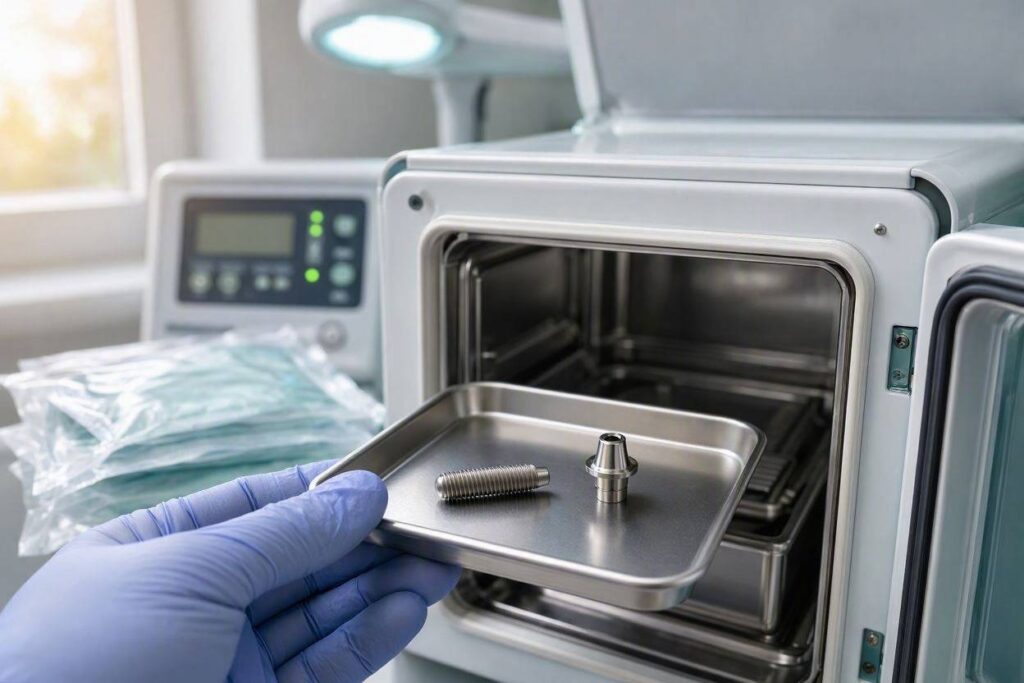
When an implant surgeon opens a small, hermetically sealed package and extracts a titanium or zirconia fixture, that device is in a state of absolute sterility. Achieving and validating this sterility is a highly regulated, rigorously documented industrial process governed by international standards, most notably ISO 11137 for radiation sterilization and ISO 17665 for moist heat sterilization.
The implant manufacturer machines the implant from raw material, cleans it to remove machining oils and debris, applies the specific surface treatment—sandblasting, acid-etching, anodization, or a combination—and then packages the implant in a specialized container designed to maintain sterility. The sealed package is then subjected to the sterilization process. For dental implants, the predominant method is gamma irradiation using Cobalt-60 as the radiation source. The ionizing radiation destroys microbial DNA, rendering bacteria, viruses, fungi, and the highly resistant bacterial spores non-viable.
Gamma irradiation is the preferred method for implants because it penetrates deeply and uniformly, does not raise the temperature of the device, does not leave toxic chemical residues, and can be applied after final packaging. The process does not alter the mechanical properties of titanium or zirconia at the dose levels used for medical device sterilization. The manufacturer validates the process by demonstrating a Sterility Assurance Level of 10 to the power of negative 6, meaning there is less than a one-in-a-million probability that a viable microorganism survives on the device.
The implant’s sterile packaging is designed with specific barrier properties. The blister pack or vial has a Tyvek lid or a similar breathable membrane that allows the sterilizing radiation to penetrate but prevents the ingress of microorganisms after sterilization. The package is sealed under controlled conditions, and the seal integrity is tested as part of quality assurance. The surgeon inspects the package before opening. If the package is damaged, wet, or the seal is broken, the implant is considered contaminated and must not be used.
What Happens the Moment Sterility Ends
The transition from sterile implant to biologically integrated prosthesis happens in seconds. The surgeon opens the sterile package, grasps the implant with sterile forceps, and places it into the prepared osteotomy site in the patient’s jaw. The implant surface is immediately bathed in blood. Blood is not sterile in the surgical sense; it contains the patient’s own commensal bacteria that enter the bloodstream from the oral mucosa, the skin, and the gingival sulcus. The surgical site, though prepared with antiseptic rinses and isolated with sterile drapes, is not a perfectly sterile field.
This is not a failure of technique. It is the biological reality of surgery in a contaminated field. The oral cavity is one of the most microbe-dense environments in the human body. The goal of sterile surgical technique is not to create a perfectly sterile wound, which is impossible in the mouth, but to minimize the bacterial inoculum to a level that the patient’s immune system can easily control. A healthy patient with a competent immune response and a well-vascularized surgical site clears the low level of bacterial contamination without clinical infection.
The blood that contacts the implant contains platelets, fibrinogen, and growth factors that immediately begin coating the titanium surface. This proteinaceous conditioning film is the foundation of healing, but it also provides receptor sites for bacterial adhesion. The race between host tissue integration and bacterial colonization begins the instant the implant is placed. In successful osseointegration, bone cells win the race, populating the implant surface and establishing a stable, bacteria-resistant interface. In peri-implantitis, bacteria win, establishing a biofilm that the host defenses cannot clear.
Peri-Implantitis: The Disease That Defies Sterilization
Peri-implantitis is the pathological condition characterized by inflammation of the peri-implant mucosa and progressive loss of supporting bone around an osseointegrated dental implant. It is a biofilm-driven disease, analogous to periodontitis around natural teeth but with important biological differences that make treatment more challenging. The question “can you sterilize a dental implant” almost always comes from a patient or clinician confronting peri-implantitis and seeking a way to salvage the failing implant.
The implant surface in peri-implantitis is colonized by a complex microbial biofilm, a structured community of bacteria embedded in a protective extracellular matrix of polysaccharides, proteins, and DNA. This biofilm adheres tenaciously to the roughened implant surface. The surface roughness that was deliberately created to promote osseointegration—the micropits and peaks that osteoblasts love—also provides ideal refuge for bacteria. Once a mature biofilm forms, it is remarkably resistant to both host immune defenses and antimicrobial agents.
The clinical challenge is that the implant threads are embedded in bone, and the infected surface extends into areas that are surgically inaccessible without reflecting a flap and removing granulation tissue. Even with surgical access, the implant surface topography makes complete debridement difficult. Bacteria lodged in the depths of the implant threads or within the micro-roughness of the surface can evade mechanical cleaning instruments. Residual bacteria repopulate the biofilm rapidly, and the disease often recurs. This is the core of the sterilization problem: you cannot sterilize a rough, threaded, partially bone-embedded device in a living human mouth.
Mechanical Debridement: The First Line of Defense
The initial phase of peri-implantitis treatment is mechanical debridement, the physical disruption and removal of the biofilm from the implant surface. This is the implant equivalent of scaling and root planing on natural teeth. The goal is to reduce the bacterial load below the threshold that triggers the host inflammatory response, allowing the peri-implant tissues to heal.
Traditional metal scalers and curettes used on natural teeth are contraindicated on titanium implants. Metal instruments scratch the titanium surface, creating new irregularities that trap bacteria and compromise the corrosion-resistant oxide layer. The standard of care is the use of instruments that are softer than titanium or non-abrasive. Plastic curettes, carbon fiber curettes, and titanium-coated curettes are designed to remove biofilm without damaging the implant surface. These instruments are used to carefully debride the exposed implant threads.
Ultrasonic scalers with specialized non-metallic tips can disrupt biofilm through acoustic streaming and cavitation. The ultrasonic energy creates microscopic bubbles in the irrigating solution that collapse and generate localized shock waves, mechanically disrupting the biofilm without requiring direct contact with the implant surface. Glycine powder air-polishing systems propel a stream of fine glycine particles in a water spray, effectively removing biofilm from the exposed implant threads with minimal surface alteration. Glycine is used because it is water-soluble and less abrasive than the sodium bicarbonate powders used on natural teeth.
The critical limitation of non-surgical mechanical debridement is access. In a deep peri-implant defect, the implant threads penetrate bone several millimeters below the gum line. The clinician can debride the supracrestal portion of the implant, the part above the bone level, but cannot reach the contaminated threads within the bony defect without surgical flap reflection. Non-surgical therapy can manage peri-implant mucositis, the reversible inflammation of the soft tissue without bone loss, but it is generally insufficient to arrest progressive peri-implantitis with established bone loss.
Chemical Decontamination and Antimicrobial Adjuncts
After mechanical debridement has disrupted the biofilm, clinicians apply chemical agents to further reduce the bacterial load and detoxify the implant surface. These agents are not sterilizing the implant; they are providing a chemical kill of residual bacteria and neutralizing inflammatory byproducts. No chemical agent applied topically in the oral environment can achieve true sterility.
Chlorhexidine gluconate is the most widely used antiseptic in peri-implantitis therapy. It is applied to the debrided implant surface either by irrigation or by placement of a chlorhexidine gel. Chlorhexidine binds to the titanium oxide layer and provides a sustained antimicrobial release over several hours. It is effective against a broad spectrum of oral bacteria. Its limitation is that it is inactivated by blood and serum, and it does not penetrate deeply into the remaining biofilm matrix.
Hydrogen peroxide, in concentrations of 3% or lower, is used for its effervescent mechanical debridement and its antimicrobial oxygen release. Tetracycline and doxycycline, applied locally as a slurry, combine antimicrobial activity with anti-collagenase effects that may stabilize the attachment of the soft tissue to the implant surface. Citric acid and EDTA gel are used for chemical detoxification of the implant surface, removing endotoxin and conditioning the titanium oxide layer for potential reattachment of soft tissue. These agents are adjuncts, not standalone solutions. They complement mechanical debridement; they do not replace it.
Laser Therapy: The Promise and the Evidence
Dental lasers, particularly the erbium:YAG and diode lasers, have been investigated for implant surface decontamination. The theoretical advantage is that laser energy can be delivered to the implant surface through a fiberoptic tip, penetrating into the depth of the defect to destroy bacteria and denature the biofilm matrix. Lasers generate localized heat, which can kill bacteria on contact, and the erbium laser’s wavelength is well-absorbed by water, making it effective at ablating biofilm without excessively heating the implant body.
The clinical evidence for lasers in peri-implantitis therapy is mixed. Some studies demonstrate superior bacterial reduction compared to mechanical debridement alone. Others show no significant difference in long-term clinical outcomes, such as probing depth reduction and bone level stability. The risk of thermal damage to the surrounding bone is real. Titanium absorbs laser energy, and the implant can heat up rapidly if the laser parameters are not precisely controlled. Temperatures exceeding 47 degrees Celsius for more than one minute can cause bone necrosis. The laser is a promising tool, but it requires specialized training and strict adherence to safe energy settings. It is not a magic wand that sterilizes a failing implant.
The Explantation Decision: When Salvage Fails
The hard clinical reality is that some peri-implantitis cases cannot be salvaged. The infection is too extensive, the bone loss is too advanced, and the implant has lost so much supporting bone that it is mobile or the defect geometry makes effective debridement impossible. In these cases, the implant must be removed, a procedure called explantation.
Explantation is not a failure of the clinician or the patient. It is the recognition of a biological limit. An implant that has lost more than 50% to 70% of its supporting bone, especially if the bone loss is circumferential rather than localized to one wall, has a poor long-term prognosis even after aggressive surgical therapy. Attempting to salvage a hopeless implant wastes time, money, and the patient’s physiological reserve. Removing the implant, grafting the defect, and allowing the site to heal before considering a new implant is often the more predictable path.
The removal of an osseointegrated implant is a surgical procedure. Specialized trephine drills, which are hollow, cylindrical burs, are used to cut a core of bone around the implant, severing the bone-implant interface. Piezoelectric surgical units use ultrasonic vibration to selectively cut bone while sparing soft tissue. The goal is to remove the implant with minimal trauma and maximal preservation of the remaining bone. The residual defect is thoroughly debrided of all granulation tissue and may be grafted with bone substitute material and a barrier membrane to prepare the site for future reimplantation.
The Reimplantation Question
After explantation and site healing, the patient and surgeon face the question of whether to place a new implant in the same location. The site that failed once has a higher risk of failing again, not because of any inherent defect in the bone, but because the patient’s host response and bacterial flora are likely unchanged. If the original implant failed due to poor oral hygiene, smoking, or uncontrolled systemic disease, those risk factors must be addressed before reimplantation. If the original failure was due to a surgical complication, such as implant placement in insufficient bone or with poor primary stability, a modified surgical approach may yield a different outcome.
The reimplantation site typically requires a healing period of four to six months after explantation and grafting before a new implant is placed. The surgeon must achieve primary stability in bone that is often less dense and less vascular than the original site. A staged approach, with a longer healing period and a submerged implant placement, is often preferred. Success rates for reimplantation are lower than for primary implant placement, but they are still acceptable in carefully selected patients.
Conclusion
A new dental implant is delivered sterile from the manufacturer via gamma irradiation, but once placed in the oral cavity, it can never be truly sterilized again due to constant exposure to the microbial environment and the impossibility of eradicating every bacterium from a biofilm-coated, bone-embedded surface. Clinical management of peri-implantitis focuses on decontamination through mechanical debridement, chemical detoxification, and laser therapy, aiming to reduce the bacterial load to a level that allows tissue health. When decontamination fails and bone loss progresses, explantation and site grafting, followed by reimplantation after healing, become the necessary salvage path.
Frequently Asked Questions
Q: Can I sterilize my implant at home with mouthwash?
A: No. Antimicrobial mouthwashes like chlorhexidine reduce the bacterial load on the accessible surfaces of the implant crown and the peri-implant sulcus, but they cannot penetrate into deeper pockets or sterilize the implant surface embedded in bone. Home care is about biofilm control, not sterilization.
Q: If an implant becomes infected, can the dentist remove it, sterilize it, and put it back in?
A: No. Once an implant is removed, it is a contaminated medical device that cannot be re-sterilized for re-use in the same patient or any other patient. The implant is discarded as biohazardous waste. A new, sterile implant is placed if reimplantation is indicated.
Q: Are some implant surfaces more resistant to biofilm than others?
A: Yes, in theory. Smoother implant surfaces, particularly at the transmucosal collar where the implant passes through the gum, accumulate less biofilm and are easier to debride. Some manufacturers have introduced hydrophilic surfaces or ultraviolet-treated surfaces that may reduce initial bacterial adhesion. However, no current implant surface is immune to biofilm formation, and the evidence for clinically significant differences in peri-implantitis rates between surface types is inconclusive.
Q: Can antibiotics cure peri-implantitis?
A: Systemic antibiotics alone cannot cure peri-implantitis. The biofilm protects bacteria from antibiotic penetration, and the avascular nature of the implant surface means antibiotics delivered through the bloodstream do not reach effective concentrations at the site. Antibiotics are an adjunct to mechanical debridement, used in aggressive or refractory cases, but they are never a standalone treatment.
Additional Resource:
For clinical guidelines on peri-implantitis management, visit the American Academy of Periodontology: https://www.perio.org/