How Do Dental Implants Fit?
You look in the mirror at your new dental implant crown. It sits among your natural teeth, and it is virtually indistinguishable from them. It does not move. It feels solid. You chew with it, you smile with it, and you clean it just like the rest of your teeth. But under the surface, a mechanical and biological marvel is at work. A titanium post is buried in your jawbone. A precisely engineered abutment connects that post to a custom-fabricated crown. How do these pieces fit together with such precision that they can withstand the crushing forces of mastication, remain stable for decades, and look like a natural part of your body? This guide will take you deep into the interface between the implant and the bone, the implant and the abutment, and the abutment and the crown. By the end, you will understand the engineering principles that make dental implants one of the most successful medical devices in history.
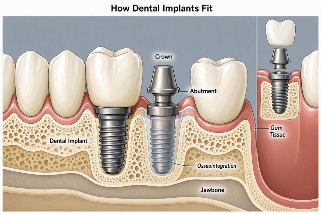
The Bone-to-Implant Interface: Osseointegration
The foundational fit of a dental implant is not a mechanical fit in the traditional sense. It is a biological bond. The implant does not simply screw into the bone like a screw into wood. It fuses with the bone at the microscopic level. This process is called osseointegration.
When the surgeon places the implant, they prepare a precisely drilled hole, called an osteotomy, in the jawbone. The diameter of this hole is slightly smaller than the diameter of the implant. The implant is screwed into the osteotomy. The threads of the implant engage the walls of the bone, creating initial mechanical stability, which is called primary stability. This is the fit you can feel immediately after surgery. It is a tight, friction-fit, screw-in-bone mechanical grip.
Over the following weeks and months, the magic of osseointegration occurs. The blood clot that forms in the microscopic gaps between the implant threads and the bone is replaced by woven bone, which then remodels into mature lamellar bone. Osteoblasts, bone-forming cells, migrate to the implant surface and deposit bone matrix directly onto the titanium oxide layer. The bone grows into the microscopic roughness of the implant surface. The interface is not a gap filled with fibrous tissue; it is a direct structural and functional connection between living bone and the implant surface. Under a microscope, the bone is intimately apposed to the titanium, with no intervening soft tissue.
This biological fit is what gives the implant its remarkable stability. The implant cannot be unscrewed without cutting the bone. It is, in essence, an ankylosed root. It has no periodontal ligament, no shock-absorbing cushion like a natural tooth. It is rigidly fixed. This is both its greatest strength, providing unmatched stability, and a factor that must be accounted for in the occlusion, the way the teeth bite together.
The Implant-Abutment Connection: Precision Engineering
The connection between the implant fixture and the abutment is a masterpiece of precision engineering. The interface must be stable, bacteria-proof, and capable of transmitting forces without loosening. Two types of connections are predominantly used.
External Hex Connection
The original Brånemark implant system used an external hexagon. The top of the implant has a hexagonal projection, typically 0.7 to 1.2 millimeters tall. The abutment has a matching hexagonal recess that fits over this projection. The external hex provides anti-rotational stability, preventing the abutment from turning, and allows the restorative dentist to position the abutment in a specific orientation. The abutment screw passes through the abutment and tightens into the internal threads of the implant, clamping the abutment down onto the implant platform. The external hex is effective but has a relatively lower resistance to lateral forces and a higher incidence of screw loosening compared to modern internal connections.
Internal Connection
Modern implant systems predominantly use internal connections. The connection is located inside the implant body. The most common designs are internal hexagons, internal octagons, and conical connections. The abutment has a corresponding male component that fits precisely into the implant’s internal female receptacle.
The conical connection, also known as a Morse taper, is a gold standard. The abutment and the implant have matching conical surfaces that are machined to incredible tolerances. When the abutment is inserted into the implant and tightened, the cones engage over a long surface area. This creates a friction-lock, a cold weld. The microscopic interlocking of the metal surfaces provides an exceptionally stable connection. The conical design also creates a bacterial seal. The tight apposition of the metal cones prevents bacteria from colonizing the internal connection and leaking out into the surrounding tissues. This is called platform switching when combined with a smaller-diameter abutment on a wider implant platform, moving the implant-abutment junction inward, away from the bone, which helps preserve bone levels.
The Fit Tolerances
The gap between the implant and the abutment is measured in microns. Machining precision of modern implants is typically in the range of 1 to 5 microns. A micron is one-thousandth of a millimeter. A gap larger than 10 microns is considered unacceptable because it allows bacterial infiltration and micromovement, which can lead to screw loosening and crestal bone loss. This is why the abutment screw is torqued to a precise specification, typically 25 to 35 Newton-centimeters, using a calibrated torque wrench. Under-tightening leaves the joint loose. Over-tightening can fracture the screw or distort the implant body. The precise torque creates the designed preload, the clamping force that maintains the stability of the connection.
The Crown-to-Abutment Fit
The final layer of fit is between the abutment and the crown. There are two primary methods of attaching the crown.
Cement-Retained Crowns
In this traditional method, the abutment is screwed to the implant. The crown is then cemented onto the abutment, much like a crown on a natural tooth. The internal surface of the crown is designed to fit the abutment precisely. A thin layer of dental cement fills the microscopic gap and provides retention.
The fit of a cemented crown must be passive, meaning it seats fully without any tension. The margin of the crown, where it meets the abutment at the gumline, must be smooth and precisely adapted. A poorly fitting margin creates an overhang that traps bacteria and cement. Excess cement, if not meticulously cleaned from the subgingival margin, is a major risk factor for peri-implantitis. This concern has driven the shift toward screw retention.
Screw-Retained Crowns
In this method, the crown has a small access channel through its biting surface. The crown is fabricated with a titanium base, a precise machined interface that fits into the implant or the abutment, and a screw passes through the crown and tightens it directly to the implant. The crown is not cemented. It is screwed into place. The screw is torqued to specification. The access hole is then filled with a tooth-colored composite resin.
The fit of a screw-retained crown is determined by the precision of the implant connection and the passive fit of the titanium base to the implant platform. There is no cement margin to worry about. The crown is retrievable. If it chips, fractures, or needs adjustment, the dentist can simply remove the resin filling, unscrew the crown, address the issue, and screw it back in. This retrievability is a major advantage.
The Role of Digital Dentistry in Precision Fit
The precision of implant fit has been revolutionized by digital dentistry. Intraoral scanners have replaced messy traditional impressions. The scanner captures a detailed three-dimensional image of the implant position, the surrounding teeth, and the soft tissue. This digital file is used to design the abutment and crown using CAD/CAM software. The restoration is then milled from a solid block of titanium, zirconia, or lithium disilicate.
The fit of a digitally milled abutment or crown is extraordinarily precise. The computer-controlled milling machine can achieve tolerances that rival or exceed those of traditional lost-wax casting techniques. Digital workflow also ensures a passive fit of implant bridges. A framework that does not fit passively can exert continuous stress on the implants, leading to bone loss and failure. Digital design and precision milling or 3D printing minimize these stresses.
The Biologic Fit: Soft Tissue Integration
Beyond the mechanical connections, the implant must fit biologically with the soft tissue. The gum tissue forms a cuff around the implant abutment, similar to the gingival cuff around a natural tooth. The epithelium attaches to the abutment surface via hemidesmosomes, creating a biological seal. This seal is critical. It prevents food debris, bacteria, and saliva from penetrating down to the bone level.
The fit of the gum tissue around the crown is the emergence profile. A well-designed emergence profile supports the gum papilla, the little triangle of gum between teeth, and creates a natural, aesthetic contour. If the emergence profile is too bulky, the gum looks swollen and unnatural. If it is too thin, the gum recedes, and the gray of the titanium abutment can show through. The fit of the implant in three dimensions, its depth, its buccal-lingual position, and its mesial-distal position, determines the quality of the soft tissue fit and the aesthetic outcome. An implant placed too far to the cheek will always look unnatural, no matter how beautiful the crown.
How the Dentist Verifies Fit
At several stages, the dentist verifies the fit of the components. After the implant is placed, primary stability is checked by assessing insertion torque and by clinical feel. After osseointegration, the implant’s stability is confirmed with a Periotest or Osstell device and by taking a radiograph to verify the absence of a radiolucent line around the implant.
When the abutment is placed, a radiograph is taken to verify that it is fully seated on the implant platform. Any gap, visible as a thin dark line between the abutment and the implant on the X-ray, indicates incomplete seating and demands correction. The crown is tried in, and the dentist uses articulating paper to verify the occlusal contacts. A thin probe is used to check the marginal fit. The patient bites down and confirms that the bite feels natural. Only then is the crown definitively tightened or cemented.
Summary of How Dental Implants Fit
- The implant fits the bone through osseointegration, a direct biological bond at the microscopic level.
- The abutment fits the implant through a precision-machined mechanical connection, with modern internal conical connections providing exceptional stability and a bacterial seal.
- The crown fits the abutment through a precisely cemented margin or a screw-retained titanium base interface.
- Digital dentistry enhances fit precision through intraoral scanning and CAD/CAM milling.
Conclusion
Dental implants fit through a seamless integration of biology and engineering. The implant fuses with the bone at the microscopic level, a living bond that anchors the restoration. The abutment connects to the implant with micron-level precision, locked in place by a calibrated screw that creates a bacteria-proof, mechanically stable joint. The crown rests on the abutment, contoured to support the gum tissue and harmonize with the natural teeth. This multi-level fit, verified at every stage with radiographs, torque wrenches, and clinical examination, is the reason dental implants can feel, function, and endure like no other tooth replacement.
Frequently Asked Questions (FAQ)
1. How tightly does the implant screw into the bone?
The implant is tightened to a specific torque during surgery, but the long-term stability comes from the bone fusing to the implant surface, not from the mechanical screw grip.
2. Is there a gap between the implant and the abutment?
In a properly fitting implant, the gap is microscopic, typically less than 5 microns, providing a bacterial seal.
3. Can an implant crown fit too tightly?
An implant crown must fit passively. A crown that is forced onto a non-passive abutment or implant can exert continuous stress on the bone, causing pain and potential bone loss.
4. Why does my implant crown not have a dark line at the gum like some crowns?
Implant crowns are made of all-ceramic materials and often use custom zirconia abutments or tissue-colored titanium bases to eliminate the dark metal shadow.
5. How does the dentist know if the implant has fused to the bone?
The dentist checks for clinical immobility, takes a radiograph to see bone contact, and may use a device like Osstell to measure implant stability quantitatively.
6. Is a screw-retained or cement-retained crown a better fit?
Both can fit equally well. Screw-retained crowns are preferred by many dentists because they eliminate the risk of retained cement and are retrievable.
7. What happens if the abutment screw loosens?
The fit of the abutment is compromised. The dentist can retighten or replace the screw. This is a manageable mechanical complication.
Additional Resource
For more information on the technology and engineering of dental implants, visit the Academy of Osseointegration:
Academy of Osseointegration