How Long Does Osseointegration Take for Dental Implant?
Osseointegration is the biological miracle upon which all of modern implant dentistry rests. The word itself, coined by Professor Per-Ingvar Brånemark, describes the direct, structural, and functional connection between living, ordered bone and the surface of a load-carrying titanium implant. It is not a chemical bond. It is a physical, microscopic interlocking and a biological acceptance. The bone cells migrate to the titanium oxide surface, attach, and begin laying down new bone matrix directly onto the treated metal, locking the implant in place with a rigidity that can withstand hundreds of pounds of chewing force. This process is silent. It happens beneath the gum, invisible to the patient. And it takes time. The question “How long does osseointegration take?” is answered not by a single number but by a range, influenced by the patient’s biology, the implant’s surface technology, and the surgical site’s bone quality. This guide is a deep, respectful exploration of the osseointegration timeline. It explains the cellular biology of bone healing around an implant, the clinical factors that compress or extend the waiting period, and the specific protocols that protect this fragile, essential process.
The Definition and Biology of Osseointegration
Osseointegration is not simply the implant becoming stuck in the bone. It is the formation of a vital, dynamic, living bone-to-implant interface without an intervening layer of fibrous connective tissue. The titanium or titanium alloy implant is not a passive scaffold. Modern implant surfaces are treated to be osteoconductive. They are sandblasted, acid-etched, and in some cases, chemically modified to be hydrophilic. This surface topography, with its microscopic peaks and valleys, is the biological invitation. It attracts blood proteins, platelets, and circulating mesenchymal stem cells.
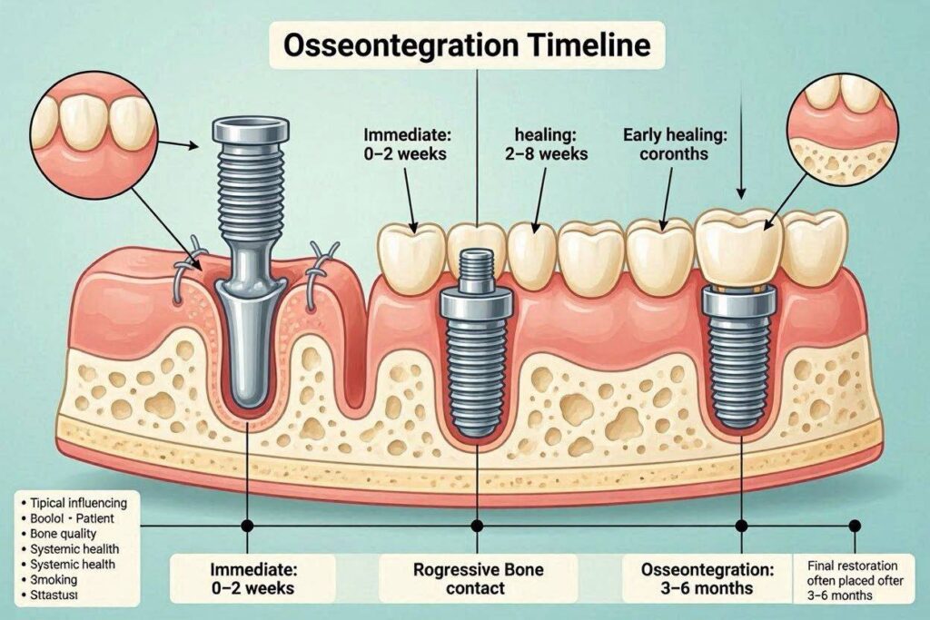
The biological sequence unfolds in stages. In the first seconds after placement, the implant surface is coated with a thin film of blood. Platelets activate and release growth factors, including platelet-derived growth factor and transforming growth factor-beta. These signals call osteoprogenitor cells to the surface. Within the first week, a provisional fibrin matrix is formed, and new blood vessels begin to sprout into the healing site, a process called angiogenesis. The osteoblasts, the bone-forming cells, then arrive. They begin secreting a collagen-rich matrix called osteoid directly onto the implant surface. This woven bone, which is immature and disorganized, is laid down rapidly. Over the following weeks and months, this woven bone is remodeled. The osteoclasts resorb it, and new, layered, highly organized lamellar bone is deposited in its place. The final, mature bone-implant interface is a rigid, continuous layer of calcified bone, in direct apposition to the titanium oxide layer. The implant is now a part of the skeleton.
The Standard Clinical Osseointegration Timeline
The clinical healing period before an implant can be safely loaded with a crown is based on decades of empirical data and histologic studies. The standard, conservative timeline is the safest for the vast majority of patients.
- Lower Jaw (Mandible): 3 to 4 months. The mandible, particularly the anterior region between the mental foramina, has dense, highly vascularized cortical and trabecular bone. The blood supply is robust, and the bone quality is typically type D1 or D2. Healing is faster and more predictable.
- Upper Jaw (Maxilla): 4 to 6 months. The posterior maxilla is composed of softer, less dense, more spongy trabecular bone, type D3 or D4. The blood supply is less abundant, and the bone is more vascular but with thinner cortical plates. Osseointegration takes longer and is more biologically demanding. If a sinus lift was performed simultaneously, the minimum wait is 6 months.
This timeline assumes a straightforward case in a healthy, non-smoking patient with good bone quality. It is a period of protected, submerged healing. The implant is buried under the gum, or a healing abutment protrudes through it, but no chewing force is applied.
The Accelerated Timeline: Modern Hydrophilic Surfaces
The premium implant manufacturers have invested heavily in surface technology to compress the osseointegration timeline. The most significant advancement is the development of chemically active, hydrophilic surfaces. The Straumann SLActive surface and the Nobel Biocare TiUnite surface are the leading examples.
A standard sandblasted and acid-etched surface is hydrophobic; it repels water and blood. A hydrophilic surface is chemically modified to be highly wettable. It actively attracts blood and serum proteins to its surface. This accelerates the initial adsorption of the fibrin matrix and the recruitment of osteoprogenitor cells. The biological consequence is a faster and more robust early bone formation. Histologic studies show a significantly higher bone-to-implant contact in the first weeks of healing.
The clinical result is a reduced healing time. Straumann documents that with the SLActive surface and a protocol of primary stability of at least 35 Ncm, the implant can be safely loaded with a temporary crown in as little as 3 to 4 weeks, even in the upper jaw. This is an accelerated, early loading protocol. It is not the standard for every case. It demands strict patient selection: excellent bone quality, high insertion torque, a non-smoker, and a controlled, light occlusal scheme. But it is a real, clinically validated option.
The Critical Factors That Influence Osseointegration Time
The timeline is not a fixed constant. It is a biological variable influenced by a matrix of patient, surgical, and implant factors.
Bone Quality and Quantity
This is the single most important anatomical variable. Dense, thick cortical bone heals with a rich blood supply and provides excellent primary stability. Soft, spongy bone heals more slowly and provides less initial mechanical grip. A site that has been heavily grafted requires extended healing for the graft to mature and revascularize before the implant can be considered integrated.
Surgical Technique and Primary Stability
An implant that is rigidly locked in place at the time of surgery, with an insertion torque of 35-50 Ncm, has the ideal mechanical environment for osseointegration. There is no micro-movement. The bone healing proceeds directly. A traumatic surgery with a dull drill that overheats the bone causes a zone of necrotic bone. Osseointegration is delayed while the body clears this dead bone, or it fails entirely.
Systemic Patient Factors
- Smoking: Nicotine is a potent vasoconstrictor. The reduced blood flow and oxygen tension in the oral tissues of a smoker dramatically slow all phases of wound healing. Osseointegration in smokers is delayed, less predictable, and has a higher failure rate.
- Diabetes: Uncontrolled hyperglycemia impairs collagen synthesis, reduces white blood cell chemotaxis, and alters microvascular circulation. Well-controlled diabetes with a low HbA1c is not a contraindication, but poorly controlled diabetes significantly compromises osseointegration.
- Osteoporosis and Medications: Osteoporosis itself is not a direct contraindication, but the bisphosphonate and RANK-ligand inhibitor medications used to treat it can severely suppress bone turnover and cause medication-related osteonecrosis of the jaw. Osseointegration is profoundly impaired.
- Age: Chronological age, in itself, does not slow bone healing. A healthy 80-year-old can osseointegrate an implant just as effectively as a 40-year-old. It is the systemic health, not the age, that matters.
The Healing Phase: What You Can and Cannot Do
During the osseointegration period, the patient has one critical responsibility: protect the implant from any force or infection. The implant site must be left undisturbed. The patient must not chew on the implant. A soft diet is mandatory for the entire healing period. The patient must maintain meticulous oral hygiene, brushing the adjacent teeth carefully and using a prescribed antimicrobial mouthwash. They must not smoke. They must not use a straw, spit forcefully, or do anything that creates negative or positive pressure in the mouth that could disrupt the wound. They must attend their scheduled follow-up appointments. The implant is a silent passenger in the bone. The patient’s compliance is a direct determinant of success.
How Osseointegration Is Confirmed
The dentist does not simply guess that osseointegration has occurred. At the end of the waiting period, the implant is uncovered. A clinical test is performed. The implant is tested for mobility. A successfully osseointegrated implant has absolutely no clinical mobility. It is rigid, immobile, like an ankylosed part of the skeleton. A periapical radiograph is taken. The X-ray should show healthy bone in direct contact with the implant threads, with no radiolucent, dark line indicating a fibrous capsule. The bone level should be at the implant platform or slightly below it. When the dentist applies reverse torque to the implant, there should be no movement and no pain. If these criteria are met, osseointegration is confirmed. The implant is ready for the prosthetic phase.
Conclusion
Osseointegration, the direct structural fusion of living bone to the titanium implant surface, takes a standard 3 to 4 months in the dense, well-vascularized lower jaw and 4 to 6 months in the softer, less dense upper jaw, with simultaneous sinus grafting extending the wait to a minimum of 6 months. Modern hydrophilic implant surfaces can accelerate this timeline to 3 to 4 weeks for carefully selected patients with excellent primary stability. The healing period is non-negotiable, protected time, and its success is directly determined by atraumatic surgical technique, rigid primary stability, and the patient’s systemic health, with smoking and uncontrolled diabetes being the most significant biological disruptors.
Frequently Asked Questions
Can I feel osseointegration happening?
No. Osseointegration is a completely silent, painless, sub-clinical biological process. You will feel no sensation of the bone fusing. The absence of pain is a good sign, but you will not feel the integration itself.
What happens if I accidentally bite on the implant during healing?
A single, light, accidental contact is unlikely to cause failure, but you must avoid any chewing force. If the implant is loaded repetitively before osseointegration, the micro-movement will cause a fibrous encapsulation instead of bone fusion. Contact your dentist if you have bitten down hard on the implant.
Can osseointegration fail after it has been confirmed?
Yes. This is late implant failure. The established osseointegration can be destroyed by peri-implantitis, a bacterial infection that resorbs the bone, or by mechanical overload that causes bone fatigue and implant fracture. Osseointegration is a living, dynamic interface that must be protected for life with meticulous hygiene.
Is there any way to speed up osseointegration naturally?
You cannot accelerate the biological rate of bone healing beyond your individual physiology. You can, however, avoid the factors that slow it down. Do not smoke, eat a protein-rich, nutrient-dense diet, control your blood sugar, and follow your surgeon’s instructions rigidly. These actions ensure you heal at your optimal biological speed.
Additional Resource:
For a deeper scientific exploration of osseointegration biology, visit the research section of the Osseointegration Foundation at www.osseo.org.
Disclaimer: The healing timelines provided in this article are general clinical guidelines. Your individual osseointegration period will be determined by your specific anatomy, medical history, and the professional judgment of your implant surgeon. Always adhere to the personalized post-operative protocol provided to you.