How to Care for Dry Grooves When Dental Implants Are Installed?
You have just completed your dental implant surgery. The numbness fades, and you follow every post-operative instruction carefully. Yet a few days later, you notice something concerning around the surgical site. The area feels different from what you expected. Perhaps you see exposed tissue, an unusual depression, or feel sensitivity where the tooth once was. Your mind immediately jumps to worst-case scenarios. Understanding what dry grooves are, how they relate to dental implants, and exactly how to care for them provides the reassurance and practical guidance needed during the critical healing period. This guide addresses the realities of post-implant tissue healing, distinguishes normal variations from concerning changes, and provides actionable care instructions that protect your investment.
Understanding Dry Grooves in the Context of Dental Implants
The term dry groove originates from post-extraction complications, specifically dry socket. However, when dental implants enter the picture, the situation changes significantly. Understanding this distinction prevents unnecessary worry while ensuring you recognize genuine problems requiring professional attention.
What Dry Socket Means in Traditional Extractions
Dry socket, medically termed alveolar osteitis, occurs after tooth extraction when the protective blood clot dislodges or dissolves prematurely. This clot serves essential functions in normal healing. It protects the underlying bone and nerve endings from oral fluids, food debris, and bacteria. It provides the scaffold for new tissue formation. It contains growth factors that stimulate healing.
When this clot is lost, the bone and nerve endings become exposed to the oral environment. The result is typically intense pain beginning three to five days after extraction, often radiating to the ear or temple. The socket appears empty or contains visible bone. A foul taste or odor may develop. This condition requires professional treatment and heals more slowly than normal extraction sites.
Dry socket occurs in approximately 2 to 5 percent of routine extractions, with higher rates for lower wisdom teeth and in smokers. Risk factors include traumatic extraction, oral contraceptive use, poor oral hygiene, and failure to follow post-operative instructions regarding spitting, straw use, and smoking.
Why Implants Change the Equation
Dental implant placement fundamentally changes the post-extraction environment. When an implant is placed immediately into an extraction socket, the implant itself occupies the space where the blood clot would form. The implant surface contacts the bony walls directly, providing the substrate for osseointegration rather than clot organization. The concept of losing a clot and exposing bone becomes less applicable because the implant fills the socket.
When implants are placed into healed ridges, there is no extraction socket at all. The implant site is prepared surgically in solid bone. A blood clot forms around the implant, but the implant provides stability and protection to the surrounding tissues. The primary closure or healing abutment further protects the surgical site.
The implant surgical site heals differently than an extraction socket. The implant-bone interface undergoes osseointegration while the overlying soft tissue heals by primary or secondary intention depending on the surgical approach. The complications that can occur are implant-specific rather than extraction-specific.
What You Might Actually Be Seeing
When patients describe a dry groove around their implant, they are often observing one of several normal or concerning findings. Distinguishing between these requires understanding what healthy healing looks like and what variations signal problems.
A healing abutment protruding through the gum creates a circular opening in the tissue that may appear as a groove or depression. This is normal and intentional. The tissue heals around the abutment, forming a cuff similar to the gum collar around natural teeth. The area should remain clean but does not require special intervention beyond gentle oral hygiene.
Gum recession around an implant can expose portions of the abutment or, in concerning cases, the implant itself. Early recession may appear as a groove or depression where tissue previously covered the site. Mild recession of 1 to 2 millimeters may be acceptable if stable. Progressive recession exposing implant threads requires evaluation and possible intervention.
Suture lines and healing incisions create temporary grooves or indentations along the gum ridge. These reflect the surgical access needed for implant placement. As healing progresses over weeks, these contours smooth and blend with surrounding tissue. The groove appearance diminishes as swelling resolves and tissue remodels.
Immediate Post-Surgical Care for Implant Sites
The first days after implant surgery determine the trajectory of healing. Proper care during this vulnerable period prevents complications and sets the stage for successful osseointegration.
The First 24 Hours: Critical Protective Measures
The initial 24 hours after implant surgery require strict adherence to protective measures. The surgical site is fresh, the blood clot is forming, and the tissues are most vulnerable to disruption. Your surgeon’s instructions for this period are not suggestions. They are requirements for protecting your investment.
Bite firmly on the gauze placed over the surgical site, maintaining pressure for 30 to 60 minutes after leaving the office. Replace gauze as directed if bleeding continues. Expect pink-tinged saliva rather than bright red bleeding after the first few hours. Do not rinse your mouth during the first 24 hours, as this disrupts clot formation.
Apply ice packs to the cheek over the surgical area in cycles of 20 minutes on and 20 minutes off. This reduces swelling and provides comfort. Keep your head elevated, even while sleeping, using extra pillows. Do not engage in physical activity that increases blood pressure, as this promotes bleeding and swelling.
Take prescribed medications exactly as directed. If antibiotics were prescribed, complete the full course even if you feel fine. Pain medication taken before the local anesthetic fully wears off provides smoother pain control than waiting until you are in significant discomfort.
Days Two Through Seven: Transitioning Care
The second day after surgery marks a transition. Bleeding should have stopped, though some oozing may persist. Swelling typically peaks around day three before beginning to subside. Bruising may appear on the cheek or under the eye for upper implants, a normal response that resolves over one to two weeks.
Warm salt water rinses begin 24 hours after surgery. Mix half a teaspoon of salt in eight ounces of warm water. Gently swish and allow the water to fall from your mouth rather than spitting forcefully. Repeat after meals and before bed. Some surgeons prescribe chlorhexidine mouth rinse instead, which should be used exactly as directed.
Oral hygiene resumes but with modifications. Brush teeth not involved in the surgical site normally. Avoid the implant area with your toothbrush for the first week unless specifically instructed otherwise. Some surgeons provide a soft surgical brush or recommend a cotton swab dipped in chlorhexidine to gently clean around healing abutments.
Diet remains soft and cool or room temperature. Yogurt, smooth soups, scrambled eggs, mashed potatoes, protein shakes, and similar foods prevent chewing trauma. Avoid hot foods and beverages, which increase bleeding. Avoid crunchy, hard, or chewy foods that could traumatize the surgical site. Do not use straws, as the suction can dislodge clots and disrupt healing.
Distinguishing Normal Healing from Problems
Healing follows predictable patterns, but individual variation exists. Understanding the range of normal reduces anxiety while ensuring you recognize when professional evaluation is needed.
Normal Post-Implant Tissue Appearance
The surgical site evolves in appearance over the first weeks. Immediately after surgery, the area appears swollen with sutures visible along the incision line. A healing abutment, if placed, protrudes through the gum as a small metal cylinder. The tissue color ranges from pink to slightly reddened due to inflammation, which is a normal part of healing.
Over the first week, swelling subsides and the tissue begins to contract around the healing abutment or cover the implant site. Sutures may begin to loosen or dissolve. A white or yellowish film may cover the healing abutment or gum surface. This is fibrin, a normal protein involved in clot formation and tissue healing, not pus or infection.
By week two, sutures have dissolved or been removed. The tissue appears pinker and less swollen. The healing abutment should be surrounded by tissue that is snug, pink, and non-tender. Some tissue shrinkage is normal as swelling resolves. The surgical contours smooth and blend with adjacent tissue.
By week four, the site should appear well-healed with mature pink tissue. A shallow groove may remain where the incision was made, but this continues to fill in over subsequent months. The healing abutment should be clean with no visible threads or implant surface below the tissue margin.
Warning Signs That Need Professional Attention
Certain signs warrant prompt communication with your surgeon. Pain that increases after day three or four, rather than improving, suggests possible infection or other complication. Pain that was well-controlled and suddenly worsens demands evaluation. Swelling that continues to increase after day three or becomes hard, hot, or spreading requires attention.
Pus or discharge from the surgical site indicates infection. A small amount of clear fluid is normal. Yellow, green, or foul-smelling drainage is not. Fever, malaise, or swollen lymph nodes accompanying local symptoms suggest systemic infection requiring urgent treatment.
Exposed implant threads or a visible increase in the amount of implant showing through the gum is concerning. While some tissue shrinkage is normal as swelling resolves, progressive exposure of the implant surface suggests tissue breakdown or bone loss. A healing abutment that becomes loose or falls out requires prompt replacement to prevent tissue closure over the implant.
Persistent numbness or altered sensation beyond 24 hours after surgery warrants evaluation. While the local anesthetic effects can linger, true nerve impairment presents as ongoing numbness, tingling, or burning in the lip, chin, or tongue. Early intervention when nerve issues are suspected improves outcomes.
Specific Care for the Tissue Around Healing Abutments
The healing abutment creates a unique environment requiring specific care approaches. This metal or ceramic cylinder extends through the gum, maintaining the tissue opening for the future restoration.
Cleaning Techniques for Exposed Abutments
Healing abutments accumulate plaque just as natural teeth do. This plaque, if not removed, causes inflammation of the surrounding tissue called peri-implant mucositis. Red, swollen, bleeding tissue around the healing abutment signals inadequate cleaning.
Gentle brushing with a soft toothbrush cleans the healing abutment effectively. Use small circular motions at the junction where the abutment meets the gum. A small amount of non-abrasive toothpaste is acceptable. Some surgeons recommend dipping the brush in chlorhexidine mouth rinse for added antimicrobial effect during the first weeks.
Interdental brushes, the small cone-shaped brushes used between natural teeth, work well for cleaning around healing abutments. Select a size that fits comfortably in the space without traumatizing the tissue. Gentle back-and-forth motion removes plaque from all surfaces of the abutment.
Water flossers on low settings can clean around healing abutments effectively, but only after the surgical site has initially healed, typically after two weeks. High pressure settings may force bacteria into the healing tissue or disrupt the forming tissue collar. Confirm with your surgeon before introducing water flossing.
Managing Food Impaction
Food debris collects around healing abutments because the tissue collar has not yet formed a tight seal. This debris causes irritation, inflammation, and unpleasant taste if not removed. Prevention and prompt removal protect the healing tissues.
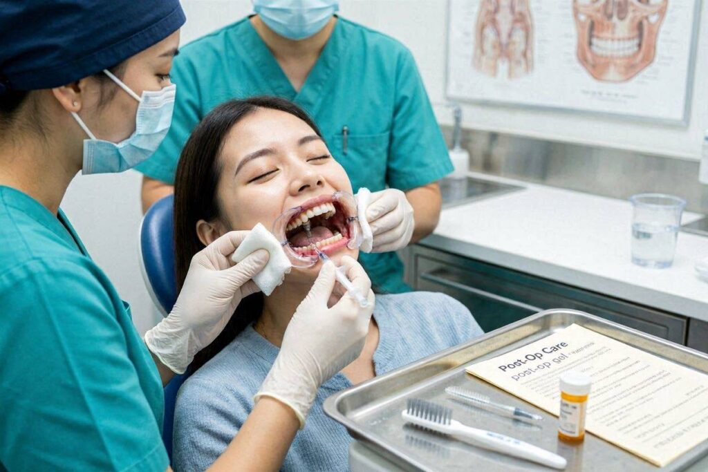
Rinsing after every meal with warm salt water or prescribed mouth rinse helps dislodge food particles before they cause problems. Gentle swishing is sufficient. A syringe with a curved tip, provided by many surgeons, allows directed irrigation of the healing abutment area. Fill with warm water or saline and gently flush around the abutment from all sides.
Do not use toothpicks or sharp instruments to remove food debris. These traumatize the healing tissue and can introduce bacteria deeper into the site. If debris remains after rinsing and gentle brushing, contact your dental office rather than attempting aggressive removal yourself.
What to Avoid Around Healing Abutments
The healing abutment screws into the implant. Excessive force can loosen it. Avoid chewing on the implant side entirely during osseointegration. This protects both the implant-bone interface and the abutment connection.
Do not play with the healing abutment with your tongue or fingers. The constant manipulation irritates tissues and can introduce bacteria. Some patients develop a habit of running their tongue over the abutment because it feels unusual. Consciously avoiding this behavior protects the healing site.
Temporary removable prostheses that contact the healing abutment require careful monitoring. The prosthesis should be relieved over the implant site to prevent pressure. Pressure transmitted through a denture or partial to the healing abutment transmits to the implant, potentially disrupting osseointegration. If you wear a temporary prosthesis, ensure your dentist has adjusted it properly and report any discomfort immediately.
Managing Complications That Mimic Dry Grooves
Several post-implant conditions produce symptoms that patients might describe as dry grooves. Understanding these conditions enables appropriate response.
Peri-Implant Mucositis
Peri-implant mucositis describes inflammation confined to the soft tissue surrounding an implant, without bone loss. This condition mirrors gingivitis around natural teeth. It is reversible with proper treatment but progresses to peri-implantitis if ignored.
The affected tissue appears red, swollen, and bleeds easily when probed or brushed. A groove or depression may be noted where inflammation has caused tissue changes. There may be discomfort but typically not the intense pain associated with dry socket.
Treatment involves professional cleaning to remove plaque and calculus from the implant surface and abutment. Specialized non-metal instruments prevent scratching the titanium surface. The patient receives reinforced oral hygiene instruction. Chlorhexidine mouth rinse may be prescribed for short-term use. With these measures, the tissue returns to health within days to weeks.
Soft Tissue Dehiscence
Dehiscence describes the opening of a previously closed surgical wound. Around dental implants, this manifests as separation of the gum tissue, exposing the underlying implant or bone. This condition most closely resembles what patients may call a dry groove.
Soft tissue dehiscence occurs for several reasons. Tension on the suture line from inadequate tissue release during surgery can pull the wound open. Pressure from a removable prosthesis can cause tissue breakdown. Infection can weaken the closure. Thin gum tissue, or lack of keratinized tissue, predisposes to dehiscence.
Small areas of dehiscence may heal by secondary intention, with new tissue gradually covering the exposed area. This process takes weeks and requires meticulous oral hygiene to prevent infection of the exposed surface. Larger dehiscence may require surgical revision to re-cover the implant. The surgeon evaluates the size, location, and cause to determine appropriate management.
Implant Thread Exposure
Exposure of implant threads through the gum represents a more serious complication. This occurs when the bone and soft tissue covering the implant resorb, revealing the roughened implant surface. Unlike a smooth healing abutment, the roughened implant surface harbors bacteria and cannot be effectively cleaned by the patient.
Early thread exposure may be managed with improved oral hygiene and professional monitoring if minimal and stable. Progressive exposure requires intervention. Surgical re-coverage with a connective tissue graft or guided bone regeneration may be attempted. In advanced cases where significant bone loss has occurred, implant removal may be necessary.
Thread exposure typically occurs on the cheek side of implants where the bone is thinnest. Prevention through adequate bone volume at placement, proper implant positioning within the bony envelope, and meticulous soft tissue handling during surgery minimizes this complication.
Tissue Management After Implant Uncovering
For two-stage implants that were buried under the gum during healing, the uncovering procedure creates a new tissue situation requiring specific care.
The Uncovering Appointment
After three to six months of submerged healing, the implant must be exposed to begin the restorative phase. This minor procedure, typically requiring only local anesthesia, locates the implant under the gum and places a healing abutment.
The surgeon makes a small incision over the implant. Any bone that grew over the cover screw is removed. The cover screw is replaced with a healing abutment that extends through the gum. Sutures may or may not be placed depending on the tissue handling. The procedure typically takes 20 to 30 minutes per implant.
Post-operative discomfort is minimal compared to the original implant surgery. Mild soreness resolves within a day or two. The healing abutment is now visible in the mouth. Tissue healing around the abutment takes two to four weeks before the restorative phase can proceed.
Care After Uncovering
The newly placed healing abutment requires the same care as one placed during initial implant surgery. Gentle cleaning, avoidance of chewing on the site, and monitoring for signs of inflammation apply. The tissue will form a collar around the abutment over several weeks.
Some bleeding from the tissue collar is normal during the first days after uncovering. The tissue was recently incised and is healing. Gentle brushing stimulates this tissue to form a tight, keratinized collar. Bleeding that persists beyond the first week or becomes heavy warrants evaluation.
The tissue collar should mature within two to four weeks. At this point, the restorative dentist takes the final impression for crown fabrication. If the tissue has not matured adequately, additional healing time is allowed before proceeding with impressions.
Long-Term Maintenance to Prevent Tissue Complications
The tissue around dental implants requires ongoing care throughout the implant’s lifetime. Preventing the development of grooves, recession, and other tissue complications depends on consistent maintenance.
Professional Maintenance Schedule
Professional implant maintenance visits should occur every three to six months, individualized based on the patient’s risk profile. These visits include evaluation of peri-implant tissue health, probing depths, bleeding on probing, and radiographic bone levels.
The dental hygienist uses instruments specifically designed for implants. These include plastic, graphite, or titanium scalers that clean the implant surface without scratching it. Scratched implant surfaces harbor more bacteria and are harder for the patient to clean. Metal instruments designed for natural teeth should never be used on implants.
Radiographs taken at appropriate intervals, typically annually, document bone levels around the implant. Stable bone levels indicate healthy peri-implant tissues. Progressive bone loss over sequential radiographs indicates peri-implantitis requiring intervention. Early detection through regular maintenance prevents advanced disease.
Home Care Tools and Techniques
Effective home care prevents the tissue inflammation that leads to recession, dehiscence, and other complications. The patient’s daily routine represents the most important factor in long-term implant health.
Interdental brushes of appropriate diameter clean the mesial and distal surfaces of implant restorations where they contact adjacent teeth. The brush should fit snugly but not tightly. Gentle back-and-forth motion cleans the abutment surface and the undersurface of the crown where plaque accumulates.
Water flossers with specialized tips for implants deliver antimicrobial solutions into the sulcus around the implant. The pulsating action disrupts plaque and flushes debris from areas mechanical cleaning cannot reach. Studies demonstrate water flosser effectiveness for reducing peri-implant inflammation.
Implant-specific floss, wider and more textured than standard floss, cleans the abutment surface effectively. Floss threaders or superfloss with stiff ends negotiate under implant-supported bridges where conventional floss cannot reach. The dental team provides instruction in proper technique for each patient’s specific restoration.
Recognizing Early Warning Signs
Patients should examine their implant restorations regularly. Changes in tissue color from pink to red or bluish indicate inflammation. Bleeding when brushing or flossing around the implant signals active peri-implant disease. Tissue swelling, tenderness, or pus on probing requires professional evaluation.
Changes in the amount of tooth structure visible may indicate tissue recession. If the implant crown appears longer than previously, or if a margin that was hidden under the gum becomes visible, recession has occurred. Early intervention when recession is first noticed may prevent progression.
Mobility of the implant restoration is always abnormal. Unlike natural teeth, which have physiological mobility, implants should be absolutely immobile. Any perceived movement warrants immediate professional evaluation. Mobility indicates either abutment screw loosening, which is treatable, or loss of osseointegration, which requires more extensive intervention.
The Role of Keratinized Tissue in Preventing Complications
What Keratinized Tissue Is and Why It Matters
Keratinized gingiva describes the firm, pale pink tissue that surrounds natural teeth and ideally surrounds implants. This specialized tissue contains keratin proteins that make it tough and resistant to mechanical injury. It attaches firmly to the underlying bone and tooth or implant surface.
Around implants, keratinized tissue provides several protective functions. It forms a tight cuff that resists bacterial penetration. It withstands the mechanical forces of chewing and oral hygiene. It remains stable over time, resisting recession. It provides the esthetic frame for the implant restoration.
Inadequate keratinized tissue around implants, typically defined as less than 2 millimeters of attached tissue, increases the risk of recession, inflammation, and patient discomfort during cleaning. Plaque accumulates more readily when the mobile, non-keratinized alveolar mucosa contacts the implant surface. Patients report more pain when brushing areas lacking keratinized tissue.
When Tissue Grafting Is Considered
Soft tissue grafting around implants may be performed before implant placement, at the time of implant surgery, at uncovering, or after restoration if problems develop. The goal is establishing an adequate zone of keratinized, attached tissue around the implant.
Free gingival grafts harvest a thin layer of tissue, typically from the palate, and transplant it to the implant site. This procedure increases the width of keratinized tissue but does not increase tissue thickness significantly. It is performed when the primary deficiency is keratinized tissue width rather than volume.
Connective tissue grafts harvest tissue from beneath the palatal surface, transplanting it to create a thicker, more resilient peri-implant soft tissue. This approach addresses both inadequate keratinized tissue and insufficient tissue thickness. Thicker tissue resists recession better and provides improved esthetics.
These grafting procedures require healing periods of six to eight weeks before the tissue is mature enough for restorative procedures. The added time investment pays dividends in long-term tissue stability and health around the implant.
Diet and Nutrition During Healing
Foods That Support Tissue Healing
Nutrition significantly influences tissue healing after implant surgery. The body requires adequate protein, vitamins, and minerals to build new tissue and fight infection. Patients who eat poorly during the healing period experience slower recovery and higher complication rates.
Protein provides the amino acids needed for collagen synthesis and tissue repair. Soft protein sources including eggs, yogurt, cottage cheese, protein shakes, and pureed meats supply these building blocks without requiring chewing. Aim for adequate protein intake throughout the healing period.
Vitamin C supports collagen formation and immune function. Citrus fruits, berries, and bell peppers supply this vitamin, though acidic foods may irritate fresh surgical sites. Supplements can bridge the gap if dietary intake is insufficient during healing. Consult your surgeon before starting supplements.
Zinc promotes wound healing and immune response. Found in meat, shellfish, legumes, and seeds, zinc deficiency impairs healing. Most patients eating a balanced diet receive adequate zinc, but supplementation may be considered for those with poor nutritional status.
Foods and Habits That Impair Healing
Certain dietary choices actively work against healing. Alcohol consumption after surgery increases bleeding, impairs immune function, and interacts with pain medications. Most surgeons recommend avoiding alcohol for at least one week after implant surgery.
Excess sugar consumption promotes inflammation and feeds oral bacteria. While the occasional sweet treat may be acceptable once initial healing has occurred, a high-sugar diet during the healing period increases infection risk and impairs tissue repair.
Smoking represents the single most damaging habit for implant healing. Nicotine constricts blood vessels, reducing oxygen delivery to healing tissues. Carbon monoxide from smoke binds to hemoglobin, further compromising oxygen transport. The chemicals in tobacco smoke directly impair osteoblast and fibroblast function. Smokers experience implant failure rates two to three times higher than non-smokers. Quitting before implant surgery and remaining smoke-free through osseointegration dramatically improves outcomes.
Comparative Overview of Post-Surgical Tissue Conditions
Distinguishing Normal from Abnormal
| Finding | Normal | Concerning | Action Required |
|---|---|---|---|
| Tissue color | Pink to slightly red | Bright red, bluish, or pale | Evaluation for inflammation or vascular compromise |
| Swelling | Peaks day 2-3, resolves by day 7 | Increases after day 3, hard or hot | Possible infection, contact surgeon |
| Healing abutment | Clean, tissue snug around it | Loose, more threads visible | Possible loosening or tissue recession |
| Sutures | Intact, loosening after day 5 | Absent with open wound before day 5 | Possible dehiscence, contact surgeon |
| Discomfort | Decreasing daily after day 3 | Increasing pain after day 3 | Possible infection or other complication |
| Drainage | Minimal clear fluid | Yellow, green, or foul discharge | Probable infection, contact surgeon |
| Bleeding | Stopped within 24 hours | Heavy or restarting after day 2 | Contact surgeon |
This table provides a framework for evaluating your healing progress. When findings fall in the concerning column, err on the side of contacting your dental office. Early intervention prevents minor issues from becoming major complications.
Tissue Changes Over the Healing Timeline
| Time Period | Expected Tissue Appearance | Expected Sensation |
|---|---|---|
| Day 1-3 | Swollen, sutures visible, some bleeding | Soreness, controlled with medication |
| Day 4-7 | Decreasing swelling, sutures loosening | Soreness diminishing |
| Week 2 | Pink, healing abutment visible | Mild tenderness on touch |
| Week 3-4 | Mature pink tissue, tissue collar forming | No significant discomfort |
| Month 2-3 | Stable tissue, implant submerged or abutment clean | Normal sensation |
| Month 4-6 | Restorative phase begins | No implant sensation |
Healing follows this general timeline, though individual variation exists. The key trend is progressive improvement. Any reversal of this trend, where symptoms worsen after improving, warrants evaluation.
When to Contact Your Surgeon Immediately
Certain post-operative developments require same-day communication with your surgical team. Pain that is not controlled by the prescribed medication regimen demands attention. Uncontrolled pain may indicate infection, nerve involvement, or other complications requiring intervention.
Excessive bleeding that does not respond to gauze pressure for 30 minutes requires evaluation. While oozing is normal, bright red bleeding that fills the mouth or persists despite pressure is not. Patients on blood thinners should have a plan established with their surgeon for managing bleeding complications.
Difficulty breathing or swallowing is a medical emergency. While extremely rare after dental implant surgery, expanding hematoma in the floor of the mouth can compromise the airway. This Ludwig’s angina-like presentation requires emergency medical attention.
Any sign of implant mobility, where the implant itself or the healing abutment feels loose, should be reported immediately. A mobile implant has not achieved or has lost osseointegration. Early recognition sometimes allows salvage procedures that late recognition does not.
Conclusion
Caring for the tissue around newly placed dental implants requires understanding that what patients describe as dry grooves typically represents either normal healing anatomy such as the healing abutment opening or suture line depressions, or manageable conditions like peri-implant mucositis or minor soft tissue dehiscence. Proper care during the critical first week includes avoiding disruption of the surgical site, maintaining gentle but effective oral hygiene around healing abutments, and adhering to soft diet restrictions that protect forming tissue. Long-term tissue health depends on consistent home care with implant-appropriate tools, regular professional maintenance, and prompt attention to early warning signs such as bleeding, color changes, or tissue recession, ensuring the implant foundation remains sound for decades.
Frequently Asked Questions
Can I get dry socket after a dental implant?
True dry socket occurs only in open extraction sockets where the blood clot is lost, exposing bone. When an implant is placed, it fills the socket, making classic dry socket unlikely. The implant surgical site can develop other complications including infection or tissue breakdown that require professional attention.
Why is there a metal post sticking through my gum?
If your implant surgery used a one-stage approach, the healing abutment protrudes through the gum intentionally. This small metal cylinder maintains the opening for the future crown and shapes the gum tissue. It is a normal part of implant treatment and remains in place until the final crown is fabricated.
What should I do if I see threads showing on my implant?
Exposed implant threads indicate tissue or bone recession and require professional evaluation. Do not attempt to cover them or ignore them. Contact your implant surgeon promptly. Early thread exposure may be manageable. Progressive exposure can lead to implant loss.
How do I clean around the healing cap without damaging anything?
Use a soft toothbrush with gentle circular motions at the junction of the cap and gum. An interdental brush or water flosser on low setting can also be effective. Avoid aggressive scrubbing. If you experience bleeding that persists beyond the first week of cleaning, consult your dental team.
Is it normal for the gum to pull away from the healing abutment?
Some tissue shrinkage is normal as swelling resolves during the first few weeks. The tissue should remain snug around the abutment. If the gap between tissue and abutment increases progressively or if you see more of the metal than you did previously, contact your dentist for evaluation.
Can I use mouthwash around my dental implant?
Alcohol-free mouthwashes are preferred, as alcohol can irritate healing tissues. Your surgeon may prescribe chlorhexidine mouth rinse for the initial healing period. After healing, non-alcohol antimicrobial rinses can supplement mechanical cleaning. Avoid mouthwashes with alcohol content during the healing phase.
How long until the groove around my implant disappears?
Suture line depressions and surgical contours typically smooth and fill in over four to eight weeks as the tissue remodels. The opening around a healing abutment remains until the final crown is placed, at which point the crown margin fills the space and the tissue adapts to the restoration contours.
What if food keeps getting stuck around the healing abutment?
Rinse after meals with warm salt water. Use a curved-tip syringe provided by your surgeon to gently irrigate around the abutment. Avoid toothpicks or sharp objects. As the tissue collar matures over several weeks, the fit around the abutment tightens and food impaction typically decreases.
Additional Resource
American Academy of Periodontology: Peri-Implant Diseases
https://www.perio.org/for-patients/periodontal-treatments-and-procedures/dental-implant-procedures/peri-implant-diseases/