What Happens When Dental Implants Fail?
The day you received your dental implant, you invested in what was supposed to be a permanent solution. You followed the healing protocol, endured the waiting period, and celebrated when your new crown was finally placed. For months or even years, everything felt fine. Then something changed. A subtle movement where there should be none. A strange sensation when you bite. A taste that was not there before. The possibility of implant failure creeps into your mind, unwelcome and unsettling. Understanding exactly what happens when dental implants fail removes the fear of the unknown and equips you with the knowledge to act promptly and appropriately. Implant failure, while uncommon, follows predictable patterns, presents recognizable signs, and offers clear treatment pathways when addressed early.
Defining Dental Implant Failure
Implant failure carries a specific clinical meaning distinct from complications that do not threaten the implant’s survival. Understanding this distinction helps patients contextualize their own situations.
What Constitutes True Implant Failure
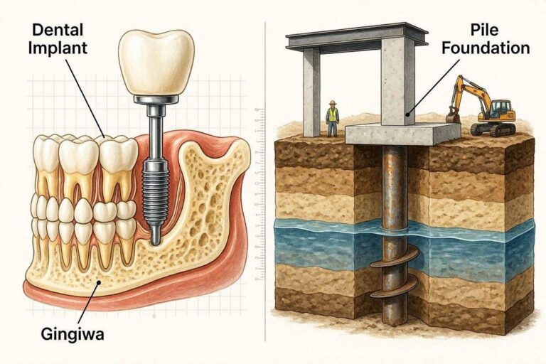
Dental implant failure means the implant has lost or never achieved osseointegration, the direct bone-to-implant contact essential for long-term function. Without osseointegration, the implant is not anchored in the bone and cannot support a functional restoration. This failure is biological rather than mechanical. The implant itself, a titanium or zirconia post, does not break down. The interface between implant and living bone fails.
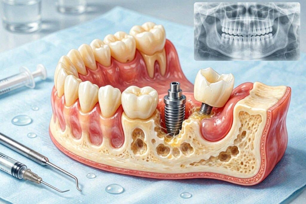
Clinically, a failed implant demonstrates mobility. Unlike natural teeth, which have a physiological ligament allowing microscopic movement, integrated implants are rigidly fixed in bone. Any perceptible movement indicates failure of osseointegration. Radiographs of failing implants show a radiolucent line, a dark space between the implant surface and surrounding bone where bone should contact the implant directly.
Implant failure differs from implant complications. A fractured abutment screw, a chipped crown, or peri-implant mucositis are problems that can be resolved without removing the implant. These complications do not constitute implant failure, though they require professional treatment to prevent progression to failure.
Early Failure vs. Late Failure
Implant failures classify by timing relative to the treatment phases. Early failures occur before the final restoration is placed, during the osseointegration period or at the time of abutment connection. These failures represent a failure of the implant to integrate with the bone.
Early failure may be discovered at the second-stage surgery when the implant is uncovered and found to be mobile. It may present during the healing period with persistent pain, swelling, or drainage that does not resolve. Radiographs taken before restoration may show failure of bone to contact the implant surface. Early failure rates in healthy patients range from 1 to 3 percent for most implant sites, somewhat higher in the posterior maxilla.
Late failures occur after the implant has been restored and in function, sometimes years after successful integration. These failures represent a breakdown of previously established osseointegration. Peri-implantitis, the inflammatory destruction of bone around an implant, is the most common cause of late failure. Overload from excessive occlusal forces can also cause late failure, particularly when combined with compromised bone support.
Late failure rates are harder to quantify because they depend on the population studied and the follow-up period. Long-term studies report cumulative late failure rates of 5 to 10 percent over ten to fifteen years, with higher rates in patients with periodontitis history, smokers, and those with poor maintenance.
Survival vs. Success: Important Distinctions
Dental literature distinguishes between implant survival and implant success. Survival means the implant remains in the mouth. Success requires the implant to meet specific criteria including absence of mobility, absence of peri-implant radiolucency, less than 0.2 millimeters of annual bone loss after the first year, and absence of pain, infection, or paresthesia.
An implant may survive but not succeed. An implant with significant bone loss, chronic inflammation, and esthetic compromise still counts as surviving if it remains in the mouth and provides some function. Patients considering implant treatment should understand that success rates, properly defined, are somewhat lower than survival rates commonly quoted in marketing materials.
The Biological Mechanism of Failure
Implant failure does not occur randomly. Specific biological processes drive the loss of osseointegration, and understanding these processes informs prevention and treatment.
Failed Osseointegration
When an implant fails to integrate, the space between the implant surface and the surrounding bone fills with fibrous connective tissue rather than bone. This fibrous encapsulation creates a soft tissue interface that cannot provide the rigid anchorage needed for implant function. The implant essentially becomes a foreign body encapsulated by scar tissue rather than an integrated part of the skeleton.
Fibrous encapsulation results from several factors. Micromovement of the implant during healing, exceeding 100 to 150 microns, directs mesenchymal stem cells toward fibroblast differentiation rather than osteoblast differentiation. The resulting tissue is fibrous rather than bony. Bacterial contamination at the time of surgery or during healing triggers an inflammatory response that favors fibrous tissue formation. Overheating the bone during surgical preparation kills osteoblasts and impairs bone formation.
Once fibrous encapsulation occurs, the implant cannot spontaneously integrate. The fibrous tissue must be removed, the site allowed to heal, and a new implant placed once the bone has regenerated. No amount of waiting will convert fibrous encapsulation to osseointegration.
Peri-Implantitis Progression
Peri-implantitis drives most late implant failures. This inflammatory condition begins with bacterial plaque accumulation on the implant surface, triggering an inflammatory response in the surrounding soft tissue. This early stage, peri-implant mucositis, is reversible with treatment. Untreated, the inflammation extends to the supporting bone, causing progressive resorption.
The bone loss around implants affected by peri-implantitis follows a different pattern than periodontitis around natural teeth. The inflammatory infiltrate around implants extends closer to the bone surface. The dense connective tissue layer that provides some protection around natural teeth is less organized around implants. As a result, peri-implantitis can progress more rapidly and may be more difficult to treat than periodontitis.
Bone loss around implants is typically circumferential, creating a crater-like defect. As bone support diminishes, the implant becomes increasingly vulnerable to occlusal forces. Eventually, the combination of bone loss and functional loading exceeds the remaining bone’s capacity, and the implant loses integration.
Overload and Biomechanical Failure
Excessive forces transmitted to the implant-bone interface can cause biological failure even in the absence of infection. This overload failure occurs when the forces applied exceed the bone’s capacity to withstand them, leading to microfractures, bone resorption, and eventual loss of integration.
Patients with bruxism, clenching, or other parafunctional habits generate forces far exceeding normal chewing loads. Without protection such as a night guard, these forces transmit directly to the implant-bone interface. The bone remodels in response, but if the forces exceed the adaptive capacity, resorption outpaces formation, and bone is lost.
Restorative factors contribute to overload. A crown with premature occlusal contacts receives forces before other teeth share the load. A cantilevered prosthesis creates leverage forces on the supporting implants. An implant placed in less-than-ideal position may receive forces at unfavorable angles. Proper restorative design and occlusal management protect against overload failure.
Immediate Consequences When an Implant Fails
The recognition of implant failure triggers a series of clinical events and patient experiences that require prompt management.
Implant Mobility and Discomfort
A failed implant moves when manipulated. This mobility may be subtle, perceived by the patient as a slight give when biting or pressing with the tongue. As failure progresses, mobility becomes obvious. The implant may rock perceptibly or even move vertically.
Pain may or may not accompany mobility. Some failed implants cause no discomfort because the fibrous tissue lacks the innervation of vital bone or the periodontal ligament. Other failed implants cause dull, aching pain, particularly under loading. Acute infection superimposed on a failing implant produces throbbing pain, swelling, and sometimes pus discharge.
The sensation of mobility in a structure that should be immovably fixed distresses patients psychologically. An implant that moves when it should be rock-solid violates expectations and generates anxiety about the implications for overall treatment. Prompt professional evaluation addresses both the physical and psychological aspects of implant failure.
Radiographic Findings
Radiographs of failed implants reveal characteristic patterns. A radiolucent line, appearing as a dark halo around all or part of the implant, indicates absence of bone-to-implant contact. This lucency may surround the entire implant or involve only a portion, typically starting at the crest and progressing apically.
Progressive bone loss on sequential radiographs confirms active failure. A single radiograph showing some bone loss may represent stable peri-implantitis. Radiographs taken months apart that show ongoing bone loss indicate progressive disease requiring intervention. The rate of bone loss provides prognostic information. Slow loss over years may be manageable. Rapid loss over months demands urgent treatment.
Cone beam CT imaging, while not used routinely for implant follow-up, provides detailed information about the three-dimensional pattern of bone loss. This information guides treatment planning when salvage procedures are being considered. The relationship of bone loss to anatomical structures such as nerves and sinuses is clearly visualized.
Functional Impairment
A failing implant cannot perform its intended function. Chewing on the affected side becomes uncomfortable or impossible. The patient unconsciously shifts chewing to the opposite side, creating asymmetric loading that can cause problems in the natural dentition or other implant restorations.
If the failing implant supported a multi-unit prosthesis such as a bridge or full-arch restoration, failure of one implant compromises the entire prosthesis. The remaining implants bear increased loads, potentially initiating a cascade of failures. Prompt management of the failing implant protects the remaining restorations.
Speech and esthetics may be affected depending on the implant location. A failing anterior implant creates visible mobility or positional change. The patient may avoid smiling or speaking clearly. The psychological impact extends beyond the physical symptoms.
The Implant Removal Procedure
When implant failure is confirmed and salvage is not possible or advisable, the implant must be removed. This procedure, while concerning to patients, is typically straightforward in experienced hands.
How Failed Implants Are Removed
The removal technique depends on the degree of implant integration and the reason for failure. A truly failed implant with no remaining osseointegration may be removed with minimal force. The surgeon grasps the implant with forceps or engages it with a removal tool and unscrews it from the bone. Because no bone remains attached to the implant surface, it rotates out with little resistance. Patients are often surprised at how quick and painless this procedure can be.
Implants that have partially integrated present more challenge. Some bone remains attached, requiring the surgeon to separate the implant from the bone. Troughing burs, thin cylindrical drills used around the implant perimeter, sever the bone-implant connection while removing minimal bone. Piezoelectric surgical devices use ultrasonic vibration to cut bone without damaging surrounding soft tissue. These techniques preserve bone for future implant placement.
In rare cases of a fractured implant, where a portion has separated and remains in the bone, removal becomes more complex. The retained fragment must be localized and retrieved, sometimes requiring a surgical window in the bone. This procedure is more invasive and results in greater bone loss than removal of an intact failed implant.
Anesthesia and Patient Comfort
Failed implant removal is typically performed under local anesthesia, the same numbing used for fillings and extractions. For anxious patients or complex removals, sedation options including oral sedation, nitrous oxide, or intravenous sedation are available. General anesthesia is rarely necessary.
Post-operative discomfort following implant removal is typically less than after the original implant surgery. The procedure is shorter, and the bone is not prepared for a new implant. Patients manage with over-the-counter pain relievers in most cases. The site heals over several weeks, with soft tissue closure occurring within two to four weeks.
Healing After Implant Removal
The implant removal site heals through a combination of blood clot organization and bone regeneration. The socket left by the implant fills with a blood clot that organizes into granulation tissue, then into new bone. This process takes three to six months depending on the defect size and the patient’s healing capacity.
During healing, the site should be protected. Patients avoid chewing on the area. Soft tissue closure occurs within weeks, but the underlying bone requires months to mature. Follow-up radiographs confirm bone fill before a new implant is considered.
The surgeon may place bone graft material into the removal site to preserve ridge dimensions for future implant placement. This socket preservation approach prevents the bone collapse that would otherwise occur during healing. The graft material serves as a scaffold for new bone formation while maintaining the ridge contours.
Bone Loss and Site Management After Failure
Implant failure leaves behind a defect that must be managed thoughtfully to enable future tooth replacement.
Assessing the Residual Bone
After implant removal and initial healing, the surgeon evaluates the residual bone quantity and quality. CBCT imaging performed several months after removal shows the bone volume available for reimplantation. The defect may have filled partially or completely, or significant bone loss may persist.
The extent of bone loss depends on several factors. The reason for original failure influences the residual bone. An implant that failed due to surgical error without infection may leave minimal bone loss. Peri-implantitis with active infection typically causes significant bone destruction that persists after implant removal. The original bone volume before the first implant was placed, the implant dimensions, and the removal technique all contribute to the residual bone.
Realistic expectations matter here. The bone after implant failure and removal rarely matches the bone that existed before the first implant was placed. Some degree of grafting is commonly required before a second implant attempt.
Grafting Protocols for Failed Implant Sites
Bone grafting of failed implant sites follows the same principles as grafting any ridge deficiency. The surgeon selects the graft material and technique based on the defect morphology and the planned future implant.
Contained defects, where the bone walls are intact, heal predictably with particulate graft materials. The walls contain the graft and provide cells for new bone formation. Non-contained defects, where one or more walls are missing, require barrier membranes to contain the graft and exclude soft tissue. Block grafts or titanium mesh may be needed for large non-contained defects.
The timing of grafting relative to implant removal varies. Some surgeons graft immediately at the time of implant removal. This approach saves a surgical procedure but risks grafting into a contaminated site if infection was present. Delayed grafting, performed after the site has healed and any infection has resolved, may be more predictable. The surgeon decides based on the clinical presentation at the time of implant removal.
Graft healing requires four to nine months, similar to primary bone grafting. Once the graft has matured, a new implant can be placed following standard protocols. The success rate for implants placed into previously failed sites after appropriate grafting approaches the success rate for primary implant placement, provided the reasons for initial failure have been addressed.
Replacement Options After Implant Failure
The failure of one implant does not preclude future implant success. Understanding replacement options helps patients make informed decisions about next steps.
Immediate vs. Delayed Replacement
Immediate replacement places a new implant at the same appointment as the failed implant removal. This approach appeals to patients who want to minimize treatment time and avoid an additional surgical procedure. However, immediate replacement carries risks.
The failed implant site may be contaminated with bacteria, particularly if peri-implantitis caused the failure. Placing a new implant into an infected site risks failure of the replacement implant. The bone defect may not be ideal for the new implant, requiring grafting that is better performed as a separate procedure. The surgeon must determine intraoperatively whether immediate replacement is appropriate.
Delayed replacement allows the site to heal completely, with or without grafting, before a new implant is placed. This staged approach takes longer but provides a more predictable environment for the replacement implant. Healing time allows resolution of any infection, maturation of graft material, and restoration of vascular supply. Most surgeons prefer delayed replacement for all but the most ideal circumstances.
Alternative Tooth Replacement If Reimplantation Is Not Possible
Some patients cannot or choose not to pursue another implant after failure. Bone loss may be too extensive for implant placement even with grafting. Medical conditions may have developed that contraindicate implant surgery. Financial considerations may limit options. The experience of failure may have reduced the patient’s willingness to undergo another surgical procedure.
Fixed bridges offer a non-surgical fixed replacement. The adjacent teeth are prepared to support a prosthesis spanning the edentulous space. This approach commits healthy tooth structure but provides good function and esthetics. Bridges typically last ten to fifteen years before requiring replacement.
Removable partial dentures replace missing teeth without surgery or tooth preparation. They cost less than fixed options but provide inferior function and comfort. Some patients find them an acceptable permanent solution, while others view them as a temporary measure until circumstances allow another implant attempt.
No replacement is also an option, particularly for posterior teeth where function and esthetics are less critical. The decision to leave a space should consider the potential for adjacent tooth drifting, opposing tooth overeruption, and the functional impact of reduced chewing surface on that side.
Factors That Predispose to Implant Failure
Understanding why implants fail enables prevention of future failures, whether of existing implants or replacement implants.
Patient-Related Risk Factors
Smoking stands as the most significant modifiable risk factor for implant failure. Smokers experience implant failure rates two to three times higher than non-smokers. The vasoconstrictive effects of nicotine reduce blood flow to healing tissues. Carbon monoxide from smoke reduces oxygen delivery. The chemicals in tobacco directly impair osteoblast and fibroblast function. Smoking cessation before implant surgery and during healing significantly improves outcomes.
Uncontrolled diabetes impairs healing and increases infection risk. Hemoglobin A1c levels above 7 percent correlate with higher implant failure rates. Well-controlled diabetes with A1c below 7 percent shows implant success rates approaching those of non-diabetic patients. Preoperative medical optimization and meticulous surgical technique improve outcomes in diabetic patients.
Periodontal disease history increases risk for peri-implantitis. The same bacterial pathogens, host response characteristics, and oral hygiene challenges that led to natural tooth loss can affect implants. Patients with periodontitis history require meticulous maintenance and more frequent professional recall to prevent implant complications.
Medications including bisphosphonates, used for osteoporosis treatment, and certain cancer therapies increase the risk of medication-related osteonecrosis of the jaw after dental surgery. This condition, characterized by non-healing exposed bone, can cause implant failure and complicate implant removal. A thorough medication history before implant surgery identifies patients at risk.
Surgical Factors
Surgical errors contribute significantly to early implant failure. Overheating the bone during implant site preparation kills osteoblasts and impairs healing. The critical temperature threshold for bone is 47 degrees Celsius maintained for one minute. Sharp drills, copious irrigation, and controlled drilling speed prevent thermal injury.
Lack of primary stability at implant placement predicts failure. The implant must be firmly anchored in bone at the time of surgery, without movement. Inadequate stability allows micromovement during healing, leading to fibrous encapsulation rather than osseointegration. The surgeon should verify primary stability and modify the plan if it is insufficient.
Bacterial contamination during surgery introduces pathogens into the sterile surgical site. Strict aseptic technique, including sterile instruments, surgical draping, and appropriate antibiotic prophylaxis when indicated, prevents surgical contamination. Perioperative chlorhexidine rinsing reduces oral bacterial load.
Implant malposition causes both biological and prosthetic problems. An implant placed too close to an adjacent tooth root can damage that tooth. An implant that perforates the buccal bone plate loses the bone support on that side. An implant angled unfavorably creates restorative challenges and non-axial loading. Careful surgical planning and execution prevent malposition.
Restorative Factors
The restorative phase can create conditions that lead to late implant failure. Excess cement left below the gum line during crown cementation is a well-documented cause of peri-implantitis. The cement itself acts as a foreign body, and the rough surface harbors bacteria. Meticulous cement cleanup or use of screw-retained restorations eliminates this risk.
Poor occlusal design subjects the implant to excessive or misdirected forces. A crown with premature contacts receives all the force before other teeth engage. A cantilevered prosthesis creates leverage that magnifies forces on the supporting implants. An implant restoration that does not allow adequate interproximal cleaning leads to plaque accumulation and peri-implant inflammation.
Inadequate fit between the abutment and implant creates a microgap where bacteria can colonize. The implant-abutment connection should be precise, with minimal gap dimensions. Original manufacturer components provide the best fit. Aftermarket or generic components may compromise this critical interface.
Psychological Impact of Implant Failure
The emotional response to implant failure deserves acknowledgment and support. Patients have invested significant time, money, and hope into the implant process. Failure feels like a betrayal of that investment.
The Emotional Response
Grief, anger, and anxiety commonly follow implant failure. The grief response may seem disproportionate for a medical device, but implants carry deep psychological significance. They represent restoration of wholeness, return of function, and investment in long-term health. Their loss triggers genuine emotional pain.
Anger may be directed at the treating clinician, particularly if the patient perceives the failure resulted from error. Even when no error occurred, the need to assign blame is a normal psychological response to adverse outcomes. Honest communication from the clinician about the reasons for failure helps process this anger constructively.
Anxiety about future treatment is natural. A patient who has experienced one implant failure may fear that subsequent attempts will also fail. This anxiety can delay needed treatment or lead to selection of less optimal options. Acknowledging and addressing this fear directly helps patients move forward.
Communicating with Your Dental Team
Open communication between patient and treatment team is essential after implant failure. The clinician should explain what happened, why it likely occurred, and what can be done to address it. Financial discussions about replacement costs and warranty coverage should be transparent.
Patients should ask questions until they understand their situation. What caused this failure? What can be done differently to prevent recurrence? What are the options moving forward, with their respective risks and benefits? What costs will be involved? A clinician who welcomes these questions and answers them thoroughly demonstrates commitment to the patient’s wellbeing.
If the patient has lost confidence in the treating clinician, seeking a second opinion is reasonable. A different perspective may confirm the original plan or offer alternatives not previously considered. The second opinion clinician needs all records, including pre-surgical imaging, surgical notes, and any post-failure imaging, to provide meaningful recommendations.
Financial Considerations After Implant Failure
The financial implications of implant failure depend on several factors, including the original treatment agreement, warranty coverage, and the replacement plan.
Warranty and Guarantee Coverage
Many implant manufacturers offer warranties on their implants. These warranties typically cover replacement of the implant fixture itself if it fails to integrate or fractures. The warranty may extend for a specific period, often a lifetime or limited number of years. However, the warranty covers only the cost of the replacement implant component, not the surgical fees or restorative costs associated with replacing it.
Some dental practices offer their own guarantees on implant treatment. These may cover re-treatment at reduced or no professional fee if failure occurs within a specified period. The terms vary widely. Patients should understand their clinician’s policy on implant failure before beginning treatment.
Dental insurance typically covers replacement of a failed implant under the same terms as the original implant, subject to annual maximums and waiting periods. If failure occurs soon after placement, the original claim may exhaust the available benefit, leaving little coverage for replacement. Understanding insurance limitations helps with financial planning.
Planning for Replacement Costs
The cost of replacing a failed implant often exceeds the original implant cost because of additional procedures required. Bone grafting, which may not have been needed initially, is often required for replacement. The total cost of implant replacement should be estimated before proceeding.
Some patients choose to delay replacement to accumulate funds or wait for insurance benefits to renew. During this delay, a temporary removable prosthesis maintains space and function. Bone grafting during implant removal preserves the site for future replacement.
Conclusion
When dental implants fail, the implant loses osseointegration, becomes mobile, and typically requires removal through a straightforward procedure performed under local anesthesia. The failure results either from failure to achieve initial bone integration due to surgical factors, patient health issues, or healing complications, or from late breakdown of established integration due to peri-implantitis or biomechanical overload. After implant removal and a healing period of three to six months, often combined with bone grafting to restore the damaged site, a replacement implant can usually be placed with success rates approaching those of primary implant placement when the causes of initial failure have been identified and addressed. The psychological and financial impacts are real but manageable through open communication with the treatment team and realistic planning for the replacement process.
Frequently Asked Questions
How do I know if my implant is failing?
Signs include mobility of the implant, pain or discomfort particularly when biting, swelling or redness of the surrounding gum, bleeding or pus, and radiographic evidence of bone loss. Any mobility in an implant is abnormal and requires immediate professional evaluation.
Can a failing implant be saved?
Some failing implants, particularly those in early stages of peri-implantitis, can be treated and maintained. Advanced bone loss or established mobility typically cannot be reversed. Early intervention when problems are first noticed provides the best chance for implant salvage.
Does implant removal hurt?
Implant removal is performed under local anesthesia, so the procedure itself is painless. Post-operative discomfort is typically less than after the original implant placement and is managed with over-the-counter pain relievers. The procedure is usually quick because the failed implant lacks bone attachment.
How long after implant removal can a new implant be placed?
The site typically requires three to six months of healing before a new implant can be placed. If bone grafting was needed, healing extends to six to nine months. Immediate replacement at the time of removal is sometimes possible but carries higher risk.
Will insurance cover replacement of a failed implant?
Dental insurance typically covers implant replacement under the same terms as the original implant, subject to annual maximums and waiting periods. Manufacturer warranties may cover the implant component cost but not surgical or restorative fees. Practice guarantees vary by provider.
What causes dental implants to fail years later?
Late implant failure most commonly results from peri-implantitis, a bacterial infection causing progressive bone loss around the implant. Excessive occlusal forces from clenching, grinding, or poor prosthetic design can also cause late failure. Medical conditions that develop after implant placement may contribute.
Can smoking cause implant failure?
Yes. Smoking is one of the strongest risk factors for implant failure, increasing failure rates two to three times. Nicotine reduces blood flow, carbon monoxide impairs oxygen delivery, and tobacco chemicals directly inhibit healing cells. Smoking cessation significantly improves implant outcomes.
What if I choose not to replace the failed implant?
Options include a fixed bridge supported by adjacent teeth, a removable partial denture, or leaving the space without replacement. Each option has functional, esthetic, and financial implications that should be discussed with your dentist to determine the best choice for your situation.
Additional Resource
American College of Prosthodontists: Dental Implant Complications
https://www.prosthodontics.org/about-acp/position-statement-dental-implants/