Is It Normal for Bone Graft Membrane to Stick Out?
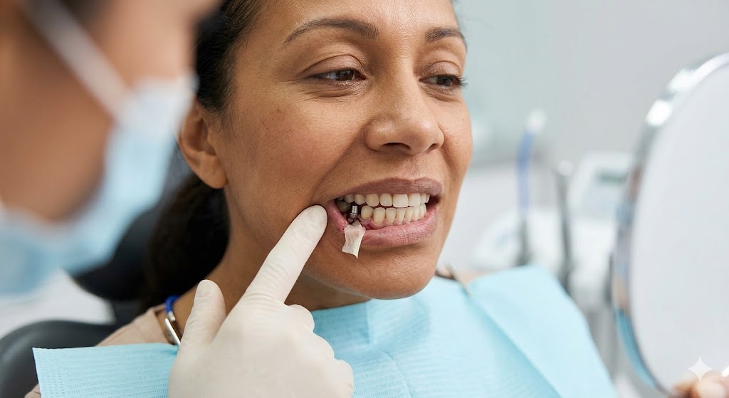
You are a few days or weeks post-surgery. You have been gently rinsing, avoiding straws, and eating soft foods. Then, one morning, you feel something strange. Your tongue touches a small, rigid, almost plastic-like edge near the surgical site. You grab a mirror, and your heart sinks. The membrane is sticking out.
Your first thought is probably panic. Did the graft fail? Did I do something wrong? Do I need emergency surgery?
Let me put your mind at ease right away. In many cases, yes, it is completely normal for a bone graft membrane to stick out. In fact, it happens so often that many oral surgeons warn patients about it before they even leave the chair.
But “normal” does not always mean “ignore it.” This article walks you through everything you need to know. You will learn why membranes expose, when to relax, and exactly when to call your dentist.
Understanding the Bone Graft Membrane: What Is It and Why Is It There?
Before we talk about exposure, we need to understand what this membrane actually is. Imagine you are landscaping a garden. You pour fresh soil (the bone graft) into a hole. If you leave it uncovered, rain (your saliva), wind (tongue movements), and animals (bacteria) will scatter the soil.
The membrane acts like a protective tarp. It is a thin, biocompatible barrier placed over the graft material.
Primary Jobs of the Membrane
- Protection: It keeps the graft particles contained so they do not migrate away.
- Exclusion: It prevents fast-growing gum tissue from invading the bone healing space. Bone grows slowly. Gum tissue grows quickly. Without a barrier, the gum would win, and you would end up with no new bone.
- Stabilization: It holds blood clots in place. Blood clots are the first step toward new bone formation.
Common Types of Membranes
Not all membranes behave the same way. Some are designed to dissolve. Others are meant to be removed. Knowing which type you have helps answer the “is it normal” question.
| Membrane Type | Material | Resorbable? | Typical Exposure Outcome |
|---|---|---|---|
| Collagen Membrane | Cow or pig collagen (purified) | Yes (dissolves in 4–8 weeks) | Very normal to see frayed, soft edges. Usually harmless. |
| PTFE (Non-resorbable) | Synthetic polymer (Gore-Tex style) | No (must be removed later) | Less common, but exposure requires a call to the surgeon. |
| High-Density PTFE | Stiffer synthetic material | No | Exposed edges can trap bacteria. Needs monitoring. |
| Synthetic Resorbable | Polyglycolic/Polyactic acid | Yes (breaks down 3–6 months) | Exposure happens but feels sharper than collagen. |
Important note: Collagen membranes are the most common today. If you see a whitish, soft, stringy or film-like piece sticking out of your gum, you likely have a resorbable membrane. This is rarely an emergency.
The Honest Answer: Yes, Exposure Is Common
Let us state this clearly. Exposure of a bone graft membrane occurs in approximately 10% to 30% of all guided bone regeneration procedures. Some studies report even higher rates for certain techniques.
Why do dentists not talk about this more? Because most exposures are small and heal without any negative effect on the graft itself.
Think of it like a scab. A scab often cracks at the edges. That does not mean the wound underneath is infected. It just means the healing process is dynamic and imperfect.
Three Main Reasons Membranes Stick Out
1. Primary Closure Failure (The Most Common Reason)
The gold standard for bone grafting is “tension-free primary closure.” That means your dentist stretches the gum flap completely over the membrane and stitches it closed without any pull.
But the mouth is a challenging environment. Swelling, talking, chewing, and even smiling create tension. If the stitches are under even a little stress, the gum can pull apart slightly. The membrane then peeks through the gap.
2. Membrane Movement or Wrinkling
During placement, membranes should lie flat. But sometimes, a small edge folds over. As swelling goes down, that folded edge pushes outward. You are not seeing the membrane migrate. You are seeing a pre-existing wrinkle that became visible when the gum tissue settled.
3. Premature Suture Dissolution or Loosening
Many oral surgeons use dissolvable stitches. These usually last 7 to 14 days. If they dissolve too early—sometimes due to acidic saliva or heavy mouth rinsing—the gum opening widens just enough for the membrane edge to slip out.
Good Exposure vs. Bad Exposure: How to Tell the Difference
Not all exposed membranes are equal. Some are harmless. Others require immediate attention. Here is a simple way to assess your situation.
Signs of a Normal, Low-Risk Exposure
- The exposed piece is small (smaller than a grain of rice or a pencil eraser).
- The membrane looks white or light tan and feels soft or slightly rubbery.
- You have no pain at the site, or only mild tenderness.
- The surrounding gum is pink (not bright red or purple).
- There is no pus, no foul taste, and no fever.
- The exposure appeared 5 to 14 days after surgery.
If your situation matches this profile, take a deep breath. You are likely fine.
Signs of a Problem Exposure (Call Your Dentist Today)
- The exposed area is growing larger day by day.
- The membrane looks black, green, or yellow.
- You feel sharp, persistent pain that painkillers do not help.
- The gum around the exposure is swollen, bleeding easily, or receding further.
- You notice a bad smell or taste coming from the site.
- You have a fever over 100.4°F (38°C) .
| Feature | Normal Exposure | Concerning Exposure |
|---|---|---|
| Size | < 3mm | > 5mm or growing |
| Color | White, tan, off-white | Black, green, dark yellow |
| Pain | None to mild | Moderate to severe |
| Gum health | Pink, firm | Red, swollen, bleeds easily |
| Discharge | None | Pus or blood |
| Systemic symptoms | None | Fever, fatigue |
Remember: Your dentist would rather receive a “false alarm” call than have you wait until an infection compromises your graft.
What Actually Happens When a Membrane Sticks Out?
Let me walk you through the biological reality. You are not broken. Your body is doing exactly what it evolved to do.
When the membrane becomes visible in your mouth, two things happen.
First, your saliva coats the exposed membrane. This is not automatically bad. Saliva has antibacterial properties. But saliva also carries oral bacteria. The longer the membrane stays exposed, the more bacteria colonize its surface.
Second, your body tries to cover the membrane again. Gum tissue has an incredible ability to creep and grow. In many small exposures, the gum will slowly grow over the exposed edge within one to two weeks. You might not even notice it happening.
However, if the exposed piece is too large or the gum is too inflamed, covering fails. That is when your dentist might need to trim the membrane or place a new stitch.
The Timeline of a Typical Minor Exposure
- Days 1–3 after exposure: You feel the edge with your tongue. Slight annoyance.
- Days 4–7: The membrane may look whiter or slightly frayed. No pain.
- Days 8–14: Gum tissue often starts climbing over the exposed edge.
- Day 15–21: Most small exposures resolve on their own as the membrane resorbs or gets covered.
Can You Still Heal Properly With an Exposed Membrane?
Yes. This is the most important takeaway.
A small, stable exposure does not automatically ruin your bone graft. The graft itself sits underneath the membrane. As long as the majority of the membrane remains intact and covered, the bone healing continues undisturbed.
Think of it like a roof with a small missing shingle. Rain might get in if the hole is large. But a tiny gap? The underlying structure stays dry.
Dental research confirms this. Studies on collagen membrane exposure show that exposures under 3mm with no infection have no statistically significant difference in bone gain compared to completely covered membranes.
When Exposure Does Cause Problems
Problems arise only when three conditions happen together:
- The exposure is large (over 5mm).
- Bacteria colonize the exposed surface.
- Inflammation spreads under the membrane to the graft particles.
When that happens, the body may start resorbing the graft prematurely. In severe cases, the graft fails completely, and you lose the bone you paid to grow.
But again, that is the minority. Most exposures never reach that stage.
Step-by-Step: What to Do If You See Membrane Sticking Out
You have looked in the mirror. You see the edge. Now what?
Step 1: Do Not Panic and Do Not Pull It
Never, ever pull the membrane. Do not grab it with tweezers. Do not cut it with scissors. Do not try to tuck it back in with a toothpick. The membrane is often anchored deep under the gum. Pulling it can tear the entire barrier, ruin the blood clot, and pull graft particles out with it.
Step 2: Assess the Size and Symptoms
Use the table above. Is this a small, white, painless exposure? Or is it large, painful, and oozing?
Step 3: Rinse Correctly
Do not use a Waterpik or a syringe pointed directly at the exposure. That high-pressure stream can lift the membrane further.
Instead, use a gentle, passive rinse:
- Mix 1 teaspoon of salt in 8 oz of warm water.
- Hold the rinse in your mouth for 30 seconds.
- Let it fall out of your mouth. Do not spit forcefully.
- Repeat 2 to 3 times per day.
Chlorhexidine mouthwash (if your dentist prescribed it) is excellent for exposed membranes. It reduces bacteria without mechanical force.
Step 4: Modify Your Diet Temporarily
For the next 7 days, avoid anything that can catch on the membrane edge:
- No chips, crackers, or crusty bread.
- No sticky foods (caramel, gummy candy).
- No seeds or small grains (rice, quinoa, poppy seeds) that can get trapped.
- Stick to smoothies, yogurt, mashed potatoes, scrambled eggs, and protein shakes.
Step 5: Call Your Dentist for These Specific Scenarios
Call the office if:
- The exposure is larger than the tip of a pencil.
- You have any pain at the site.
- You see swelling spreading to your cheek or jaw.
- The exposure happened after a sneeze, cough, or heavy exercise (sudden pressure changes can enlarge exposures).
- You are a smoker (smokers have higher graft failure rates with exposures).
Do not wait for your next scheduled appointment if any of the above apply.
Professional Treatments for Exposed Membranes
If you see your dentist and they decide intervention is needed, do not worry. The solutions are usually quick and minimally uncomfortable.
Option 1: Observation With Chlorhexidine
For very small, asymptomatic exposures, the dentist may simply prescribe a medicated mouthwash and schedule a follow-up in one week. No physical treatment is performed.
Option 2: Membrane Trimming
The dentist numbs the area with topical gel or a tiny injection. Then, using sterile scissors or a scalpel, they trim the exposed portion of the membrane. This removes the bacteria-trapping surface. The remaining membrane stays under the gum. The gum often heals over the trimmed edge within days.
Option 3: Re-suturing
If the gum opening is wide, the dentist may place one or two new stitches to pull the gum closed again. This is more common with non-resorbable PTFE membranes.
Option 4: Membrane Removal (Rare)
In cases of severe infection or graft failure, the dentist may remove the entire membrane. This is a last resort. The graft may still survive without the membrane if the bone particles have already started fusing.
Common Myths About Bone Graft Membrane Exposure
Let me clear up some misinformation that floats around dental forums and social media.
Myth 1: “Any exposure means the graft failed.”
False. As we covered, 10-30% of grafts have some exposure. The vast majority succeed.
Myth 2: “You should rinse aggressively to clean the membrane.”
False. Aggressive rinsing lifts the membrane further. Gentle rinsing only.
Myth 3: “Collagen membranes turn into goo and get absorbed by the gum.”
Partially true. They resorb, but they do not dissolve like sugar. They break down into fragments that your body removes. Some fragments may work their way out through the gum. That is also normal.
Myth 4: “If you see the membrane, you need antibiotics.”
False. Antibiotics treat bacterial infection, not the presence of the membrane itself. Unnecessary antibiotics promote resistance. Only take them if your dentist diagnoses an infection.
Myth 5: “Exposed membranes always smell bad.”
False. A mild, neutral odor is normal. A putrid, rotting smell is a red flag.
How to Prevent Membrane Exposure Before It Happens
Prevention is always easier than treatment. If you are scheduled for a bone graft, or you are in the early days of healing, follow these guidelines.
Choose an Experienced Surgeon
Not all dentists place membranes with the same skill. Ask potential surgeons:
- How many guided bone regeneration procedures do you perform per month?
- What is your membrane exposure rate?
- Do you use tension-free closure techniques?
A honest surgeon will tell you exposures happen but should be rare for large exposures.
Follow Post-Op Instructions Religiously
This sounds obvious, but here is what patients actually do wrong:
- Smoking: Even one cigarette dramatically reduces blood flow to the gum tissue. That blood flow is what heals the gum over the membrane. If you smoke, your exposure risk triples.
- Using a straw: The suction pulls on the blood clot and can lift the membrane edge.
- Chewing on the opposite side: Yes, you know this. But many patients forget when they are hungry and distracted. The graft side must have zero chewing pressure for at least two weeks.
- Touching the site with your tongue: This is a hard habit to break. But constant tongue pressure pushes the membrane outward. Consciously rest your tongue on the roof of your mouth.
Ask About Stitch Type
Non-dissolvable stitches hold longer. Some surgeons use them specifically for patients at higher risk of exposure (thin gum tissue, smokers, large grafts). The downside? You need a second appointment to remove them.
Long-Term Healing: What Happens to the Membrane Over Time?
Understanding the full lifecycle of the membrane helps you stay calm when you see changes.
Week 1-2: The membrane is intact, white or off-white. It acts as a rigid barrier. Exposures, if they happen, usually occur during this period due to suture loosening or swelling resolution.
Week 3-6: If you have a resorbable collagen membrane, it starts breaking down. It may look fuzzy, frayed, or develop small holes. This is resorption, not failure. The body is digesting the collagen.
Week 7-12: The membrane is mostly gone. Only small fragments may remain. The new bone underneath is now solid enough to survive without the barrier.
Month 4-6: No membrane remains. The gum tissue has completely healed. The bone graft is now integrated into your jaw. You cannot see or feel any remnants.
If you have a non-resorbable PTFE membrane, your dentist will schedule a second surgery to remove it. This is usually done at 4 to 6 months. Do not assume it will dissolve on its own.
Special Scenarios and Higher-Risk Situations
Not all patients have the same risk profile for membrane exposure. Here are specific cases where you need extra vigilance.
Thin Gum Biotype (Thin Gingiva)
Some people naturally have thin, almost translucent gum tissue. If you can see the outline of your tooth roots through your gums, you likely have thin biotype. These patients have much higher exposure rates. Ask your dentist about using a thicker membrane or a two-layer technique.
Multiple Adjacent Grafts
If you received bone grafts for several teeth in a row (e.g., teeth 8, 9, and 10), the gum flap is larger and under more tension. Exposure at one of the incision lines is more likely.
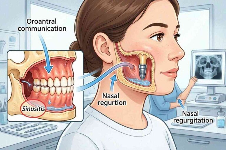
Sinus Lift With Membrane
A sinus lift is a different procedure, but it often uses a membrane too. Sinus membrane exposures are less common visually because you cannot see them. But if you feel air or fluid passing from your nose to your mouth, call your dentist immediately.
Immunocompromised Patients
If you have diabetes (especially uncontrolled), autoimmune disease, or take steroids or biologics, your healing is slower. Even small exposures can become problematic. Your dentist may recommend prophylactic antibiotics or more frequent follow-ups.
Frequently Asked Questions (FAQ)
Q1: How long does it take for an exposed membrane to heal on its own?
A: For small exposures under 3mm with no infection, most heal or get covered by gum tissue within 2 to 4 weeks. The membrane itself may resorb completely in 6 to 8 weeks.
Q2: Can I brush my teeth near the exposed membrane?
A: Yes, but with extreme care. Use an extra-soft toothbrush. Do not brush directly over the exposure. Brush the teeth adjacent to the site using gentle circular motions. Avoid the gumline near the exposure entirely.
Q3: Will the exposed membrane affect my dental implant placement later?
A: Usually not. As long as the bone graft heals with adequate volume and density, a small membrane exposure does not prevent future implant placement. Your surgeon will remove any remaining membrane fragments during implant surgery.
Q4: My membrane turned black. What does that mean?
A: Black discoloration often indicates necrotic (dead) tissue or heavy bacterial colonization. Call your dentist today. Do not wait for your next appointment.
Q5: Can I use hydrogen peroxide on the exposed membrane?
A: No. Hydrogen peroxide is too harsh for healing gum tissue. It can damage the edges of the gum and delay closure. Stick to salt water or prescribed chlorhexidine.
Q6: Is it normal for the membrane to smell?
A: A very mild, almost metallic or “medical” smell can be normal as the membrane breaks down. A strong, foul, rotting odor suggests infection. Call your dentist if the smell is offensive.
Q7: My membrane fell out completely. What now?
A: Do not panic. If the graft was placed 4 or more weeks ago, the bone may already be stable enough. If it happened in the first two weeks, call your dentist. They may need to place a new membrane. Do not throw the membrane away—save it in a clean bag for your dentist to see.
Q8: Will my insurance cover membrane removal if needed?
A: Most dental insurance plans cover necessary membrane management (trimming, removal) as part of the original bone graft procedure. However, if the exposure was caused by poor home care or smoking, some plans may deny coverage. Check with your provider.
Realistic Expectations: Your Healing Roadmap
Let me give you a week-by-week guide so you know exactly what to expect, exposed membrane or not.
Week 1
- What you feel: Swelling, mild to moderate pain, stitches.
- Membrane status: Fully intact, completely covered by gum in ideal cases.
- If exposed: You may notice a tiny white dot or line near the stitches.
Week 2
- What you feel: Swelling down, stitches dissolving or loosening.
- Membrane status: May start showing at the edges as gum shrinks.
- If exposed: The exposed area may grow slightly as swelling resolves.
Week 3
- What you feel: Almost normal, but the site is still tender to pressure.
- Membrane status: Collagen membranes begin softening.
- If exposed: The exposed edge may look frayed. This is resorption, not failure.
Week 4
- What you feel: You forget about the graft most of the day.
- Membrane status: Partially resorbed. Non-resorbable membranes still intact.
- If exposed: By now, most small exposures are either covered or clearly stable.
Week 6
- What you feel: Normal function, but avoid heavy chewing on that side.
- Membrane status: Collagen membranes largely gone.
- If exposed: Exposed collagen fragments may still be visible but are breaking down.
Month 3
- What you feel: Completely normal. You may forget which side had the graft.
- Membrane status: No membrane remains for resorbable types.
- If exposed: Any previous exposure site should be healed.
Month 6
- What you feel: Ready for implant placement (if that was the plan).
- Membrane status: Gone.
- If exposed: No evidence of prior exposure remains on exam.
Summary Table: Do I Call My Dentist or Wait?
| Symptom | Action |
|---|---|
| Small white edge, no pain | Wait, monitor daily |
| Small white edge, mild tenderness | Call during office hours |
| Large exposed area (>5mm) | Call today |
| Black or green membrane | Call immediately |
| Pus or bleeding | Call today |
| Fever over 100.4°F | Call immediately or go to urgent care |
| Foul smell + pain | Call today |
| Membrane fell out completely | Call during office hours |
A Note From the Author
You are doing the right thing by seeking information. Dental surgery recovery is stressful. The mouth is highly sensitive, and every small change feels alarming.
But here is the truth I wish every patient knew: Minor membrane exposure is a technical complication, not a disaster. Most of the time, it does not change your final outcome. You will still get the bone you need. You will still get your dental implant. You will still smile with confidence.
Do not let anxiety convince you that you ruined your graft. You did not.
Be vigilant. Be smart. Rinse gently. Call your dentist when you are unsure. And trust the process. Your body knows how to heal, even when the path looks a little messy.
Additional Resource
For a visual guide and more patient stories about bone graft healing, visit the American Association of Oral and Maxillofacial Surgeons (AAOMS) patient education page:
Link: https://myoms.org/procedures/bone-grafting/
Note: This link leads to a trusted professional organization. Always verify any online information with your personal dentist.
Conclusion
Seeing a bone graft membrane stick out of your gum is understandably frightening, but in most cases it is a normal, manageable event. Small, painless, white exposures typically heal without harming the graft, while large, painful, or discolored exposures require a call to your dentist. The key is to stay calm, avoid touching or pulling the membrane, and follow the simple rinsing and dietary guidelines until your dentist can evaluate the site.
Disclaimer: This article is for informational purposes only and does not constitute medical or dental advice. Every patient’s anatomy, healing capacity, and surgical procedure are unique. Do not use this information to diagnose or treat yourself. Always consult your own oral surgeon or dentist for specific guidance about your bone graft recovery. If you are experiencing severe pain, fever, or difficulty breathing, seek emergency medical care immediately.