What Are the Symptoms of Dental Implant Failure?
You run your tongue over the implant crown for the hundredth time, trying to decide if what you feel is real or imagined. There is a sensation that was not there before. Maybe a slight give when you press on it. Maybe the gum looks different than it did last month. Maybe there is a taste you cannot quite identify. You search online, but the symptoms listed seem vague, overlapping with normal healing variations and minor issues. You do not want to be the patient who rushes to the dentist over nothing, but you also know that ignoring early warning signs can turn a manageable problem into a lost implant. This guide provides a comprehensive, realistic framework for recognizing the symptoms of dental implant failure, distinguishing them from benign conditions, and understanding exactly when professional evaluation becomes urgent.
Understanding Implant Failure Symptoms
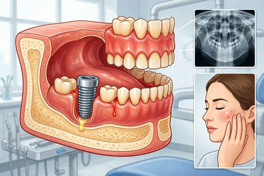
Implant failure does not announce itself with a single unmistakable sign in most cases. Instead, it presents through a constellation of symptoms that develop over time. Recognizing these symptoms early, understanding their significance, and acting promptly can mean the difference between saving an implant and losing it.
Why Early Recognition Matters
The window for intervention in implant failure is narrow but real. Peri-implant mucositis, the reversible precursor to peri-implantitis, can be treated and resolved when caught early. Once bone loss becomes established, treatment shifts from cure to management. By the time an implant becomes mobile, salvage is rarely possible.
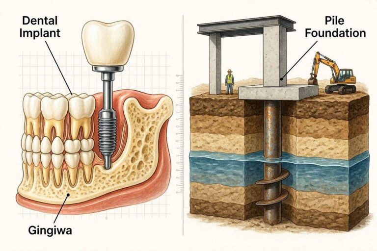
Implants lack the protective mechanisms of natural teeth. Natural teeth have a periodontal ligament that provides sensory feedback about overload and a dense connective tissue barrier that slows bacterial invasion. Implants have neither. Inflammation spreads faster. Bone loss progresses more rapidly. The absence of early warning pain, which natural teeth provide through the periodontal ligament, means patients may not notice problems until significant damage has occurred.
Regular professional monitoring provides the safety net that implants themselves lack. The combination of patient awareness of symptoms and scheduled professional evaluation creates the best defense against undetected implant failure.
The Spectrum from Minor Issues to Major Failure
Implant-related symptoms exist on a continuum. At one end are benign, self-limiting issues related to normal healing or minor soft tissue irritation. At the other end is catastrophic failure with implant mobility, gross infection, and significant bone loss. Between these extremes lies a progression where early intervention changes outcomes.
Understanding where your symptoms fall on this spectrum requires honest assessment and professional correlation. A single symptom in isolation may be insignificant. Multiple symptoms occurring together paint a more concerning picture. Symptoms that persist or worsen despite home care warrant evaluation. Symptoms that resolve with improved hygiene and professional cleaning suggest manageable soft tissue inflammation rather than true implant failure.
Pain and Discomfort Patterns
Pain serves as the body’s alarm system, but its relationship to implant failure is nuanced. Some failing implants cause significant pain, while others cause none until advanced damage has occurred.
Pain Quality and Timing
The quality of implant-related pain provides diagnostic clues. Dull, aching pain that persists or worsens over time suggests inflammation or infection in the bone surrounding the implant. This pain may be constant or may intensify when biting or tapping on the implant. It differs from the sharp, transient sensitivity of natural teeth because implants lack the nerve supply that creates that specific sensation.
Pain on biting or loading is particularly significant. An integrated implant should transmit forces to the bone without discomfort. Pain when chewing on the implant crown suggests inflammation at the bone-implant interface or, in advanced cases, movement of the implant within the bone. This symptom always warrants professional evaluation.
Night pain or pain that awakens the patient from sleep raises concern for significant infection or nerve involvement. Unlike daytime pain that may be triggered by function, spontaneous nocturnal pain suggests an ongoing pathological process that does not require mechanical stimulation to produce symptoms.
Distinguishing Surgical Pain from Failure Pain
Normal post-surgical pain follows a predictable timeline. Discomfort peaks within the first 48 to 72 hours after implant placement, then steadily decreases. By one week post-surgery, most patients require little or no pain medication. By two weeks, the surgical site should be essentially comfortable, though mild tenderness on firm palpation may persist.
Pain that deviates from this timeline requires attention. Pain that increases after day three or four, rather than decreasing, suggests possible infection. Pain that was well-controlled and suddenly worsens after a week or more of improvement raises concern. Pain that persists at a significant level beyond two weeks post-surgery warrants evaluation.
Late-onset pain, developing months or years after successful implant integration and restoration, is never normal. Such pain indicates a new problem, most commonly peri-implantitis, overload, or mechanical failure of the implant components. The implant that was comfortable for years and becomes painful requires the same thorough evaluation as a natural tooth with new-onset pain.
When Pain Is Absent Despite Failure
Some of the most advanced implant failures present with no pain at all. The implant may be mobile, the surrounding bone may be significantly resorbed, and infection may be present, yet the patient feels nothing. This phenomenon occurs because the implant-bone interface lacks the sensory innervation of the periodontal ligament.
Patients with gradually progressive peri-implantitis may be unaware of their condition until a clinician identifies it during a maintenance visit. The bone loss advances silently. The soft tissue inflammation may produce mild bleeding on brushing that the patient dismisses as normal. By the time pain develops, if it ever does, the condition may be far advanced.
This silent progression makes regular professional evaluation essential for implant patients. The maintenance visit, including probing, bleeding assessment, and periodic radiographs, detects problems the patient cannot feel. Skipping these visits risks allowing treatable conditions to progress to untreatable ones.
Mobility: The Definitive Sign
Mobility represents the most definitive and concerning symptom of implant failure. An integrated dental implant is rigidly fixed in bone through osseointegration. Any perceptible movement means that osseointegration has failed or is failing.
Types of Mobility
Different types of mobility indicate different problems. True implant mobility, where the entire implant moves within the bone, indicates loss of osseointegration. This movement may be rotational, where the implant can be slightly turned, or lateral, where the implant rocks side to side. Either type indicates the implant is no longer bone-anchored and cannot support a functional restoration.
Abutment mobility, where the abutment or crown moves but the implant body remains stable, indicates a mechanical rather than biological problem. The abutment screw may have loosened or fractured. This condition, while requiring prompt treatment to prevent damage to the implant internal connection, does not represent implant failure. Tightening or replacing the abutment screw typically resolves the issue.
Prosthesis mobility may involve the crown moving on the abutment due to cement failure or, for screw-retained restorations, the entire crown-abutment complex moving due to abutment screw loosening. Again, this mechanical problem is fully reversible with appropriate treatment and does not threaten the implant itself.
Self-Testing and Its Limitations
Patients commonly discover implant mobility by pressing on the crown with a finger or tongue and perceiving movement. This discovery is often distressing, as the expectation is absolute rigidity. The patient should not repeatedly manipulate a mobile implant, as this can further damage the compromised bone-implant interface.
Formal mobility testing is performed by the clinician using instrument handles to apply controlled force in different directions. The clinician distinguishes between implant mobility, abutment mobility, and prosthesis mobility, which the patient may not be able to differentiate. Radiographs confirm the clinical findings and assess the extent of bone loss associated with the mobile implant.
If you suspect implant mobility, seek professional evaluation promptly. Do not delay hoping the problem will resolve spontaneously. It will not. A mobile implant will only become more mobile over time as the bone continues to resorb. Early removal of a failed implant preserves bone for future replacement.
Perceived vs. Actual Mobility
Some patients perceive mobility that is not actually present. The normal physiological mobility of adjacent natural teeth, which move slightly within their periodontal ligaments, can create the illusion that the implant is moving when the teeth are used as a reference point. The implant is rigid, but the tooth moves, making the implant feel like it moved.
Similarly, a loose temporary restoration or a removable prosthesis that rocks on the implant can feel like implant mobility when the implant itself is stable. A dentist can quickly differentiate perceived from actual mobility through examination. Do not panic based on perceived mobility, but do seek confirmation through professional evaluation.
Gum and Soft Tissue Changes
The soft tissue surrounding an implant provides visible evidence of the underlying health or disease state. Changes in this tissue are often the earliest patient-observable signs of developing problems.
Color Changes and Inflammation
Healthy peri-implant tissue appears pink and firm, similar to healthy gum around natural teeth. There may be slight variation in pigmentation based on the patient’s ethnic background, but the tissue should be uniform in color and texture around the implant.
Redness indicates inflammation. The tissue may appear bright red or dusky red, depending on the chronicity of the inflammation. Redness confined to the marginal tissue, the 1 to 2 millimeters immediately adjacent to the implant, suggests early peri-implant mucositis. More diffuse redness extending several millimeters from the implant margin suggests more established inflammation.
Bluish or purplish discoloration may indicate venous congestion associated with chronic inflammation. This color change, sometimes subtle, suggests that the inflammatory process has been present for some time. Pale, whitish tissue may indicate fibrosis or, if combined with a rolled margin, may suggest the tissue has adapted to chronic low-grade inflammation.
Swelling accompanies inflammation. The tissue loses its firm, knife-edged contour and becomes puffy, rounded, and enlarged. Swelling may be confined to the interdental papilla, the triangular tissue between the implant and adjacent tooth, or may involve the entire circumference of the implant. Edema, or fluid swelling, indicates active inflammation.
Bleeding Patterns and Significance
Bleeding on gentle probing or brushing is the most sensitive early indicator of peri-implant inflammation. Healthy peri-implant tissue does not bleed with normal oral hygiene or gentle professional probing. Bleeding indicates ulceration of the sulcular epithelium, the thin tissue lining the crevice between the implant and the gum, which is a hallmark of inflammation.
The pattern of bleeding provides information. Bleeding confined to a single location may indicate a local irritant such as retained cement, an overcontoured crown margin, or a specific oral hygiene challenge. Circumferential bleeding, around the entire implant, suggests more generalized peri-implant inflammation.
Bleeding severity correlates roughly with inflammation severity. A few drops of blood that stop quickly suggest mild mucositis. Profuse bleeding that continues after the stimulus is removed suggests more significant inflammation. Spontaneous bleeding, occurring without any manipulation, indicates severe inflammation with tissue fragility.
A critical distinction: bleeding does not necessarily mean the implant is failing. Peri-implant mucositis, the reversible inflammatory condition, produces bleeding. The absence of bone loss distinguishes mucositis from peri-implantitis. However, persistent bleeding that does not resolve with improved oral hygiene within two to three weeks warrants professional evaluation to rule out progression to peri-implantitis.
Pus and Suppuration
Purulent discharge, or pus, from around an implant is an unequivocal sign of infection. Unlike bleeding, which may indicate reversible mucositis, pus indicates an established infectious process that has overwhelmed the tissue’s immune defenses.
Suppuration may be obvious, with visible pus exuding from the peri-implant sulcus spontaneously or when pressure is applied. It may be subtle, detected only when the clinician probes the sulcus and sees a small amount of cloudy or yellow fluid. The patient may notice a persistent bad taste or odor that does not resolve with oral hygiene.
The presence of pus means the implant is infected. The infection may be confined to the soft tissue, or it may extend to the bone. Radiographic evaluation determines whether bone loss has occurred. Implants with suppuration require prompt professional treatment including mechanical debridement, antimicrobial therapy, and possibly surgical access for thorough cleaning of the implant surface.
Recession and Tissue Loss
Gum recession around an implant exposes structures that should be covered. Mild recession of 1 to 2 millimeters may be stable and acceptable, particularly if the exposed surface is the smooth abutment or crown margin. Progressive recession that continues over time or exposes implant threads is concerning.
Recession exposes the implant surface, which is roughened to promote osseointegration. This roughened surface, once exposed to the oral environment, accumulates plaque and calculus that patients cannot effectively remove. The resulting chronic inflammation drives further tissue and bone loss, creating a vicious cycle.
Tissue loss may present as a visible increase in the length of the implant crown. The patient notices that more of the crown is visible than before, or that the gum line no longer matches the adjacent teeth. This is often the first sign of recession, appearing before the implant surface itself becomes visible.
Radiographic Signs
Radiographs provide the definitive assessment of bone support around implants. While patients cannot take their own radiographs, understanding what their dentist is looking for helps them participate meaningfully in monitoring their implant health.
What Healthy Implant Radiographs Show
A healthy implant radiograph shows the implant body surrounded by bone without any dark lines or gaps. The bone level should be at or near the first thread of the implant. Slight crestal bone loss of 1 to 1.5 millimeters during the first year after restoration is considered normal remodeling. After this initial remodeling, bone levels should remain stable over time.
The bone surrounding the implant should appear uniform in density, without areas of decreased radiodensity that would indicate bone loss. The implant threads should be clearly visible, surrounded by bone that appears in close contact with the implant surface. The crestal bone should form a smooth, continuous line from the adjacent tooth to the implant.
Comparison with previous radiographs is essential. A single radiograph provides a snapshot. Serial radiographs taken over time reveal trends. Stable bone levels over multiple years indicate a healthy implant. Progressive bone loss over sequential images indicates active disease.
Patterns of Pathologic Bone Loss
Peri-implant bone loss appears on radiographs as a radiolucent, or dark, area around the implant where bone should be. This lucency may take several patterns depending on the underlying cause.
Horizontal bone loss proceeds evenly around the implant, with the bone level reducing uniformly on all sides. This pattern typically indicates plaque-induced peri-implantitis. The bone loss may be shallow or deep, but it maintains a relatively flat contour around the implant.
Vertical or angular bone loss creates a defect on one side of the implant, with bone remaining at a higher level on the other side. This pattern may indicate overload on the affected side, a localized irritant such as retained cement, or an anatomical factor that predisposed that specific area to bone loss.
A circumferential radiolucent line surrounding the entire implant, separating it from the bone, indicates complete loss of osseointegration. This is the radiographic hallmark of a failed implant. The implant may appear to be floating in a dark halo, with no bone contact visible anywhere along its surface.
Furcation involvement, where bone loss extends between adjacent implants, indicates breakdown of the interimplant bone. This pattern is particularly concerning because the blood supply to this bone is limited, making regeneration difficult once it is lost.
Frequency of Radiographic Monitoring
The American Academy of Periodontology and other implant organizations recommend radiographic evaluation at implant placement, at abutment connection, at final prosthesis delivery, and at regular intervals thereafter. Annual radiographs are typical for stable implants, though patients with risk factors may require more frequent imaging.
Patients should understand that radiographs are not taken at every maintenance visit unless there is a specific indication. The radiation exposure, while minimal with digital radiography, should be justified by clinical need. Changes in probing depths, bleeding patterns, or patient symptoms may trigger radiographic evaluation between scheduled intervals.
Changes in Function
Implants exist to restore function. Changes in how the implant functions, or how it feels during function, can signal developing problems that may not yet be visible or painful.
Altered Sensation When Chewing
A properly integrated implant transmits chewing forces efficiently and without sensation. The patient should be unaware of the implant as a separate entity. The tooth feels like part of the jaw, not a foreign object.
A sensation of pressure, movement, or give when biting on the implant suggests a problem. This may be subtle initially, described as a springy feeling or a sensation that the tooth depresses slightly under load. As bone loss progresses, the sensation becomes more pronounced. The patient may describe feeling as though the tooth is moving or that something is not right when chewing on that side.
Audible or palpable clicking when biting can indicate several issues. The abutment screw may be loose, creating a clicking as the crown-abutment complex moves on the implant. The cement seal between crown and abutment may have failed, allowing movement. Less commonly, the implant itself may be moving within the bone, creating a duller sound or sensation.
Food Impaction Changes
An increase in food trapping around an implant that previously did not trap food suggests a change in the tissue contours or the contact relationship with the adjacent tooth. This symptom, while seemingly minor, can indicate progressive tissue changes or bone loss.
The open contact between implant crown and adjacent tooth allows food to pack vertically during chewing. This impaction traumatizes the interdental papilla, causing inflammation and discomfort. The persistent food debris contributes to bacterial accumulation and peri-implant inflammation.
Food impaction that develops long after restoration delivery may indicate tooth movement. Natural teeth drift over time, potentially opening a contact that was initially tight. The implant, rigidly fixed, cannot drift to maintain contact. The result is a food trap that worsens over time unless the restoration is adjusted or replaced.
Thermal Sensitivity
Natural teeth respond to hot and cold through fluid movement in the dentinal tubules, stimulating nerve endings. Dental implants lack this mechanism because they have no natural tooth structure, no dentin, and no nerve supply within the tooth.
True thermal sensitivity should not occur with implants. If a patient experiences cold or hot sensitivity associated with an implant, the sensation likely originates from an adjacent natural tooth. The brain localizes sensation imprecisely, and a sensitive adjacent tooth may be perceived as coming from the implant.
Persistent sensitivity to hot or cold that the patient attributes to the implant should be evaluated, with careful testing to identify the actual source. The implant itself is not the source, but the evaluation may reveal problems with adjacent teeth or with the implant’s soft tissue health that require treatment.
Systemic Symptoms
Implant failure typically remains localized to the implant site. However, significant infections can produce systemic symptoms that the patient should recognize.
Fever and Malaise
Fever in association with an implant is concerning for significant infection. Localized peri-implantitis rarely causes fever. An abscess associated with a failing implant, where pus accumulates under pressure, can produce systemic symptoms including fever, chills, and general malaise.
Swollen lymph nodes under the jaw or in the neck indicate that the immune system is responding to a localized infection. Tender, enlarged nodes on the same side as the implant suggest that the infection has spread beyond the immediate implant site. This finding warrants prompt evaluation and likely antibiotic therapy.
These systemic symptoms are not subtle. The patient feels unwell in a way that clearly differs from their baseline. Fever is measurable, not just a subjective feeling of warmth. When systemic symptoms accompany implant-related complaints, urgent evaluation is indicated.
Sinus Complications with Upper Implants
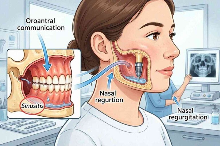
Implants placed in the posterior maxilla lie in close proximity to the maxillary sinus. When these implants fail or develop peri-implantitis, the infection can extend into the sinus cavity, causing sinusitis.
Symptoms of implant-related sinusitis include unilateral sinus pressure, congestion on the side of the implant, post-nasal drip, and sometimes foul-smelling nasal discharge. These symptoms may be mistaken for an ordinary sinus infection, delaying recognition of the implant as the source.
Sinus communication, where a passage opens between the mouth and the sinus through the implant site, can occur with failing upper implants. The patient may notice air passing between the mouth and nose, fluid leaking from the nose when drinking, or a bubbling sensation in the implant area when blowing the nose. This complication requires surgical repair in addition to managing the failed implant.
Taste Disturbances
A persistent bad taste that localizes to the implant area suggests infection. The taste may be metallic, sour, or frankly foul. It does not resolve with brushing, mouthwash, or dietary changes. The taste may intensify when pressure is applied to the implant or surrounding tissue.
The source of the taste is typically purulent drainage from the peri-implant sulcus. Even small amounts of pus that are not visible can produce a noticeable taste. The patient may also notice halitosis that does not respond to normal oral hygiene measures.
While taste disturbances can have many causes, including medications, sinus conditions, and dietary factors, a taste that localizes specifically to an implant site should prompt evaluation for peri-implant infection.
Timeline of Symptom Development
Implant failure symptoms develop along recognizable timelines that correlate with the underlying pathological process. Understanding these timelines helps patients and clinicians interpret symptoms appropriately.
Early Failure Symptoms
Early implant failure, occurring during the osseointegration period before restoration, presents with symptoms that overlap with normal post-surgical healing. Distinguishing failure from normal healing requires attention to the trajectory of symptoms over time.
Persistent pain beyond the expected post-operative period is the most common early failure symptom. Instead of decreasing after the first week, pain continues or worsens. The surgical site remains tender, and the patient continues to require pain medication beyond the prescribed course.
Swelling and redness that do not resolve according to the normal healing timeline suggest infection or failed healing. The tissue around the implant may appear boggy, inflamed, and friable rather than progressing to pink and firm. Sutures may dehisce, with wound edges separating to expose the underlying implant.
Radiographs taken when early failure is suspected show failure of bone to integrate with the implant surface. Instead of the bone density increasing around the implant as healing progresses, a radiolucent line persists or develops. The implant may be mobile when tested, though clinicians avoid loading implants during the healing period specifically to protect forming osseointegration.
Late Failure Symptoms
Late failure, occurring after successful restoration and a period of function, most commonly presents with soft tissue changes noticed by the patient or clinician before pain or mobility develops.
Bleeding on brushing or flossing around the implant, particularly if this is a new development, is often the first sign. The patient may notice pink in the toothpaste when brushing or blood when using interdental cleaning aids. This bleeding represents peri-implant mucositis that, if untreated, can progress to peri-implantitis.
Gradual changes in the appearance of the gum around the implant follow. The tissue may appear redder, more swollen, or may begin to recede. The patient may notice that the implant crown appears longer than before, or that the gum line no longer matches adjacent teeth. These changes occur slowly over months or years and may be attributed to aging or normal variation.
Deepening of the peri-implant sulcus, detected during professional probing, indicates attachment loss. While the patient cannot measure this at home, they may notice that food traps more readily, that floss goes deeper than before, or that the tissue feels different when probed with the tongue.
Pain is a late symptom of peri-implantitis, often not appearing until significant bone loss has occurred. The pain is typically dull and aching, worse with biting, and reflects inflammation in the bone rather than the implant itself. By the time pain develops, the condition may be advanced.
The Speed of Progression
The rate at which implant failure symptoms progress varies based on the underlying cause and patient factors. Peri-implantitis in a smoker with poor oral hygiene may progress rapidly, with significant bone loss occurring over months. The same condition in a non-smoker with good hygiene may progress slowly over years.
Overload failure can progress quickly once bone loss begins. The implant-bone interface, already compromised, receives forces that further damage the remaining bone, accelerating the failure process. An implant that was stable for years can become mobile within months if overload is significant and unaddressed.
Mechanical problems like abutment screw loosening develop gradually, with the screw loosening progressively until the crown movement becomes noticeable. The timeline from initial loosening to patient awareness varies based on occlusal forces, screw design, and whether the patient attends regular maintenance where screw torque can be checked.
Self-Assessment and Decision-Making
Patients must decide when symptoms warrant professional attention. A systematic approach to self-assessment aids appropriate decision-making.
Questions to Ask Yourself
When evaluating a potential implant problem, systematically consider these questions. Am I experiencing pain, and if so, is it constant or only when biting? Is the pain improving or worsening over time? Does the implant feel different than it did previously? Is there any movement I can detect? Do the gums around the implant look different in color, shape, or position? Is there bleeding when I clean around the implant? Is there any discharge or bad taste?
Answering these questions creates a clearer picture to communicate to the dental team. A patient who reports that their implant feels slightly different and the gums bleed when they floss but there is no pain or mobility provides useful information that guides the urgency and focus of the evaluation.
When to Monitor at Home vs. When to Call
Minor, isolated symptoms of short duration may be appropriate for home monitoring with improved oral hygiene. Slight bleeding that appears after a lapse in flossing and resolves with resumed cleaning suggests reversible mucositis. Mild tissue redness that improves with better brushing technique is likely benign.
Symptoms that persist beyond two weeks despite improved home care warrant professional evaluation. Any mobility, even slight or intermittent, requires prompt attention. Pain, particularly pain on biting, should be evaluated. Pus or discharge should never be ignored. Changes that are progressive, where the situation is clearly getting worse, need professional intervention.
When in doubt, err on the side of contacting your dental office. A phone call describing your symptoms allows the clinical team to triage appropriately. They may recommend home care measures with a follow-up timeline, or they may schedule an evaluation based on the specific symptoms described. The cost of an unnecessary visit is far less than the cost of a lost implant.
Differential Diagnosis: Symptoms That Mimic Failure
Several conditions produce symptoms that can be confused with implant failure. Accurate diagnosis prevents unnecessary treatment and anxiety.
Peri-Implant Mucositis
Peri-implant mucositis produces redness, swelling, and bleeding around the implant without bone loss. These are the same soft tissue signs that accompany early peri-implantitis. Only radiographic evaluation distinguishes between the two conditions.
Mucositis is fully reversible with professional cleaning and improved home care. The tissue returns to health within days to weeks. The implant is not failing. However, untreated mucositis is the precursor to peri-implantitis, so these symptoms should not be ignored even though they do not currently threaten the implant.
Retained Cement Peri-Implantitis
Cement-retained implant restorations risk leaving excess cement below the gum line. This cement acts as a foreign body, triggering inflammation that mimics plaque-induced peri-implantitis. The symptoms are identical: bleeding, swelling, bone loss on radiographs, and sometimes suppuration.
This condition is iatrogenic, meaning caused by the treatment itself. The cement must be removed, often requiring surgical access, for the tissue to heal. Once the cement is removed, the bone defect may regenerate spontaneously or may require grafting. The implant itself is not failing, but the cement-induced inflammation will cause failure if not addressed.
Mechanical Complications Without Biological Failure
Loose abutment screws produce a mobile crown on a stable implant. The patient perceives mobility and may assume the implant is failing. The treatment is simple: access the screw, retighten it to the specified torque, and reseal the access hole. The implant is not compromised.
Fractured abutment screws or abutments produce similar symptoms but require component replacement rather than simple retightening. The implant body remains integrated. Once the damaged component is replaced, function is restored.
Porcelain fracture on the implant crown creates sharp edges, altered bite, and esthetic compromise. While distressing, this is purely a restorative problem. The implant itself is unaffected. The crown may be repaired or replaced without disturbing the implant.
Adjacent Tooth Pathology
Pain perceived as coming from the implant may actually originate from an adjacent natural tooth. Tooth decay, cracks, or pulp inflammation in a neighboring tooth can create referred pain that the brain localizes to the implant.
The clinician systematically tests all teeth in the quadrant to identify the true source of pain. Percussion testing, thermal testing, and radiographs of adjacent teeth complement the implant evaluation. Finding and treating the actual problem in the adjacent tooth resolves the perceived implant symptoms.
Symptom Documentation for Your Dental Visit
Effective communication with your dental team improves diagnostic accuracy and treatment efficiency. Preparing for your evaluation appointment ensures you convey all relevant information.
What to Track Before Your Appointment
Note the timeline of your symptoms. When did you first notice something was different? Has the situation remained stable, improved, or worsened? Are there specific triggers that provoke symptoms, such as biting, hot or cold foods, or certain oral hygiene activities?
Describe the quality of any pain: dull, sharp, throbbing, constant, intermittent. Rate the severity on a scale of one to ten to provide an objective reference point. Note any home care measures you have tried and whether they helped.
Take a photograph if visible changes are present. A smartphone photo of the implant and surrounding gum, taken in good light, provides documentation that can be compared to the clinical appearance at the time of your visit. Symptoms that come and go may not be visible during the appointment, but your photograph captures them.
Questions to Ask Your Dentist
Prepare questions to ask during your evaluation. Is this a soft tissue problem or a bone problem? Has there been bone loss since my last radiograph? Can this implant be saved, or does it need to be removed? What are the treatment options, and what are their success rates? What caused this problem, and how can we prevent recurrence? What is the timeline for treatment and healing?
Write these questions down before your appointment. Anxiety during the visit can cause you to forget concerns that seemed clear at home. A prepared list ensures you leave the appointment with the information you need.
Conclusion
The symptoms of dental implant failure span a spectrum from subtle soft tissue changes like bleeding, redness, and swelling, through progressive signs like recession, pus, and radiographic bone loss, to definitive evidence including implant mobility and pain on function. Early recognition of peri-implant mucositis, the reversible precursor condition, provides a critical window for intervention before irreversible bone loss occurs. Implant mobility represents the definitive sign of failure, indicating loss of osseointegration that cannot spontaneously recover and typically requires implant removal. Regular professional monitoring with probing, bleeding assessment, and periodic radiographs detects changes the patient cannot perceive, as many failing implants progress silently without pain until advanced damage has occurred. Any symptom that persists beyond two weeks of improved home care, any mobility, any suppuration, or any pain associated with an implant warrants professional evaluation to distinguish between manageable complications and true implant failure requiring removal and replacement.
Frequently Asked Questions
How can I tell if my dental implant is failing?
The most common early signs are bleeding when brushing or flossing around the implant, redness or swelling of the surrounding gum, and gradual gum recession. More serious signs include persistent pain, especially when biting, any perceived movement of the implant, and pus or discharge. Implant mobility is the definitive sign of failure and requires immediate professional attention.
Is some bleeding around implants normal?
No. Healthy peri-implant tissue does not bleed with brushing, flossing, or professional probing. Bleeding indicates inflammation, either reversible peri-implant mucositis or more serious peri-implantitis. Intermittent bleeding that resolves with improved hygiene may be acceptable, but persistent bleeding warrants professional evaluation.
Can a failing implant hurt even years after placement?
Yes. Late implant failure from peri-implantitis or overload can cause pain years after successful integration and function. Pain that develops in a previously comfortable implant is never normal and should be evaluated. The pain is typically dull and aching, often worse with biting.
What does a loose implant feel like?
An implant that has lost osseointegration may feel like it gives slightly when pressed or when biting. There may be a rocking sensation or the feeling that the implant moves independently of the surrounding bone. Any perceived movement in a structure that should be absolutely rigid is concerning.
How quickly does implant failure progress?
The speed varies. Peri-implantitis can progress slowly over years or rapidly over months, depending on the bacterial virulence, host response, and risk factors like smoking. Overload failure can progress quickly once bone loss begins. Early failure during healing typically becomes apparent within weeks.
Can antibiotics fix a failing implant?
Antibiotics alone cannot resolve peri-implantitis because they do not remove the bacterial biofilm from the implant surface. Mechanical debridement of the contaminated implant surface is essential. Antibiotics may be used as an adjunct to mechanical treatment. True implant failure with loss of osseointegration cannot be reversed with antibiotics.
Are there warning signs I can see in the mirror?
Yes. Look for redness or swelling of the gum around the implant, recession that makes the implant crown appear longer, and any exposed metal that was previously covered by gum tissue. Compare the implant site to the surrounding teeth and to previous photographs if available. Changes in color, contour, or tissue position warrant evaluation.
What should I do if I think my implant is failing?
Contact your implant dentist or a dental specialist promptly. Describe your symptoms clearly. Do not delay hoping the problem resolves spontaneously, as it typically will not. Early intervention provides the best chance for saving a compromised implant or, if it cannot be saved, preserving bone for a successful replacement implant.
Additional Resource
American Academy of Implant Dentistry: Dental Implant Complications
https://www.aaid.com/patient_education/dental_implant_complications.html