What Does Mfl Mean In Dentistry?
In dental charting and clinical notation, MFL is an abbreviation that stands for “Mesial Fissure Lingual.” It is a specific topographic descriptor used to pinpoint the exact location of a carious lesion or a restoration on the occlusal surface of a tooth. Understanding this three-letter code requires a foundational knowledge of dental anatomy and the standardized naming conventions that allow every dental professional to communicate a precise, three-dimensional location on a tooth without ambiguity.
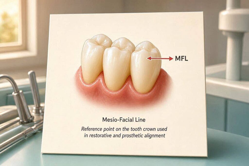
Decoding the Anatomical Terminology
Every tooth surface has a named direction. The dental profession uses a universal vocabulary derived from Latin roots to describe these surfaces. “Mesial” means the surface of the tooth that faces toward the midline of the dental arch. On a posterior tooth, the mesial surface is the side that touches the tooth in front of it. “Distal” is the opposite, facing away from the midline and toward the back of the mouth.
“Fissure” is a specific anatomical feature, not a surface. A fissure is a deep, narrow groove or cleft in the enamel of the occlusal, or chewing, surface of a premolar or molar. These developmental grooves are created when the cusps of the tooth fuse together during formation. Fissures are the most susceptible sites on a tooth for developing decay because they trap food debris and plaque, and a toothbrush bristle is often too wide to clean them effectively.
The Lingual Surface Defined
“Lingual” is the surface of a tooth that faces the tongue. On a lower posterior tooth, the lingual surface is the entire inner wall of the tooth. On an upper posterior tooth, this same surface is anatomically more correct to call the palatal surface, as it faces the palate. However, the abbreviation “L” is used almost universally for “lingual” in shorthand charting for both upper and lower teeth, with some systems using “P” for palatal on upper teeth.
Therefore, when a dentist writes “MFL” on a tooth diagram, they are not describing a random spot. They are describing a specific, defined point. The lesion or filling is located at the end of a fissure line, where that groove descends down the mesial aspect of the tooth, specifically on the lingual side of that mesial marginal ridge.
The Clinical Picture of an MFL Lesion
To visualize an MFL caries or restoration, picture a lower first molar. Look down at its biting surface. You will see a complex pattern of grooves, the central fissure system. Follow the main groove forward, toward the front of the mouth. It ends at the mesial marginal ridge, a raised rim of enamel that forms the front wall of the tooth. Just before it crosses this ridge and becomes a smooth surface contact with the tooth in front, the groove splits or curves. The part that veers toward the tongue side of the marginal ridge is the mesial fissure lingual point.
A filling described as an “MFL composite” or an “MFL amalgam” is a small, precisely placed restoration. It occupies this specific corner of the occlusal surface. It is more extensive than a simple pit-and-fissure sealant, but it is not a large, multi-surface restoration. The dentist has removed the decay at this specific vulnerable point and restored it, leaving the rest of the healthy fissure system intact if possible, or the filling may extend slightly into the central groove.
MFL vs. Other Fissure Abbreviations
The MFL code is part of a family of fissure location abbreviations. The dentist may also chart a DFL (Distal Fissure Lingual), which is the mirror-image point on the back side of the tooth, where the fissure meets the distal marginal ridge on the lingual side. An MFB (Mesial Fissure Buccal) describes the same mesial point but on the cheek-facing side of the marginal ridge. A DFB (Distal Fissure Buccal) completes the set of four corner fissure locations.
These codes are distinct from the standard surface codes used for billing. A typical billing code for a restoration is described by the number of surfaces it covers: MO (mesial-occlusal), DO (distal-occlusal), MOD (mesial-occlusal-distal). An MFL restoration is a specific subset of an occlusal restoration. For billing purposes, it is usually recorded as a one-surface occlusal filling or as part of an MO filling if the decay extends into the mesial surface. The MFL notation is the purely clinical, anatomical descriptor; the MO or O is the billing and laboratory code.
The Diagnostic Process for MFL Caries
Detecting decay at an MFL point is a daily diagnostic challenge in clinical practice. The dentist uses a combination of visual, tactile, and radiographic methods to find and confirm a lesion in this hidden, shadowy corner of the tooth. The process is methodical and critical, as a missed MFL cavity will progress silently into the dentin.
The first step is visual inspection with a dry, well-illuminated tooth. The dentist aims a stream of compressed air across the fissure system to clear it of saliva. A chalky, opaque white discoloration or a dark brown to black staining directly at the MFL point is suspicious. A dental explorer, a fine, sharp instrument with a curved tip, is then very gently moved across the fissure. A sticky, tugging sensation as the tip catches in a soft area of demineralized enamel is a strong clinical sign of cavitated decay. Magnification loupes with a bright headlight are standard of care for this fine, detailed inspection.
Radiographic Confirmation
A bitewing radiograph is essential. The X-ray beam is directed perpendicular to the tooth and the film, providing a clear view of the interproximal surfaces and the occlusal enamel. On the radiograph, the dentist looks for a dark, triangular radiolucency at the MFL point, just beneath the mesial marginal ridge on the lingual side. Early MFL decay is often invisible on an X-ray because it is hidden within the thick enamel of the cuspal slopes. More advanced decay that has penetrated the dentin appears as a classic, dark shadow that spreads laterally at the dentinoenamel junction. The clinical suspicion and the radiographic shadow together form the diagnosis that leads to restorative treatment.
The Restorative Treatment
Restoring an MFL lesion is a common, conservative dental procedure. The goal is to remove the diseased tissue and seal the site without weakening the tooth unnecessarily. The dentist anesthetizes the tooth, isolates it with a rubber dam, and uses a small, round bur in the high-speed handpiece to access the MFL decay precisely. The cavity preparation is minimal. Only the infected enamel and soft, leathery dentin are removed. Because the MFL point is at the margin of the tooth, the preparation must be carefully shaped to protect the marginal ridge from fracture.
The cavity is restored with a bonded, tooth-colored composite resin. The dentist uses a total-etch or self-etch bonding protocol to create a micromechanical and chemical bond between the composite and the remaining tooth structure. The composite is placed in small increments, each one light-cured for a precise time. The final restoration is contoured to replicate the original MFL fissure anatomy, polished to a high shine, and checked with articulating paper to ensure the bite is perfectly comfortable. The result is a small, invisible, and highly functional restoration that preserves almost all of the healthy tooth structure.
Conclusion
MFL in dentistry is the abbreviation for Mesial Fissure Lingual, a precise anatomical landmark on the occlusal surface of a posterior tooth where a groove meets the mesial marginal ridge on the tongue-facing side. This highly specific notation guides the dentist’s diagnostic focus and the restorative treatment plan. An MFL composite or amalgam filling represents conservative, preventive dentistry at its finest: removing a small, early cavity at a predictable, vulnerable pit and preserving the maximum amount of healthy tooth structure for a lifetime.
Frequently Asked Questions
Q: Is an MFL filling the same as an MO filling?
A: Not exactly. An MFL is a very specific spot on the occlusal surface, at the mesial fissure on the lingual side. If the decay extends down onto the smooth mesial surface, breaking through the marginal ridge, the filling becomes a two-surface mesial-occlusal restoration. The MFL notation describes the precise location within the fissure system.
Q: Why does the dentist use these complicated abbreviations instead of plain English?
A: These abbreviations are a universal, standardized clinical language. When a dentist reads a chart from a colleague or a specialist, “MFL” instantly and unambiguously communicates the exact location on the tooth, eliminating any possible confusion that a long descriptive phrase might cause.
Q: Can an MFL cavity be sealed with a sealant instead of a filling?
A: If the MFL point is just a deep, clean, stain-free fissure, a preventive resin sealant is an excellent, non-invasive option. If there is any staining, stickiness, or radiographic evidence of decay into the dentin, a filling is necessary to remove the infected tissue.
Additional Resource
For a complete and illustrated guide to dental anatomy, tooth numbering systems, and the precise surface and fissure nomenclature used in clinical charting, consult the official student and practitioner resources.
- American Dental Association Dental Anatomy Resources: ada.org