Where Does Cadaver Bone for Dental Implants Come From?
A dental implant is a modern marvel, a testament to the possibility of restoring function and a natural-looking appearance to a smile compromised by tooth loss. However, the foundation of a successful implant is not just the titanium post itself. It is the volume and health of the jawbone that surrounds it. When a tooth is lost, the body, in its remarkable but often unhelpful efficiency, begins to resorb the bone that once held that tooth. It decides the bone is no longer needed and reclaims its minerals. For many patients, this leaves a jawbone too thin or too shallow to securely house an implant. This is where a bone graft becomes the essential, unsung hero of implant dentistry. Your dentist might mention the need for a graft, and then add a phrase that gives many patients pause: “We can use cadaver bone.” The mental image is jarring and raises a host of profound and entirely valid questions. Where does this bone come from? Is it safe? Is it ethical? This guide is a deep, respectful exploration into the world of cadaver bone for dental implants—a world of rigorous science, stringent regulation, and a gift that transforms lives.
Defining the Term: Allograft Bone
The clinical term for cadaver bone is an “allograft.” In medical terminology, a graft is a tissue transplant. An “autograft” is tissue moved from one site to another in the same individual, such as taking bone from your hip to place in your jaw. An “allograft” is a tissue transplant between two genetically non-identical members of the same species—from one human to another. This is distinct from a “xenograft,” which is a tissue transplant from one species to another, like bovine (cow) or porcine (pig) bone, which are also commonly used in dentistry. The term “cadaver bone” is stark and clinical. The term “human donor tissue” speaks more clearly to the reality: this is a medical gift, given in the wake of loss, to heal another person. The use of human allograft bone in dentistry is a specialized branch of a much larger field of transplant medicine, governed by an extraordinarily rigorous set of tissue banking standards.
The Origin: The Gift of Donation
The journey of cadaver bone for your dental implant does not begin in a laboratory. It begins with a profound and conscious act of human generosity—the decision to be an organ and tissue donor.
The Donor Profile and Consent Process
Donors are individuals who have passed away, and whose medical and social history can be thoroughly screened. Unlike organ donation, which requires a beating heart, tissue donation can occur up to 24 hours after cardiac death, provided the body has been kept cool. Every donor or their legal next-of-kin must provide explicit, detailed consent. A generic organ donor designation on a driver’s license is a powerful first step, but tissue donation typically requires a separate, more specific authorization process, often facilitated by a trained coordinator from a tissue bank. The family is approached with immense sensitivity. They are provided with clear information that the bone tissue will be used in medical procedures to help recipients heal, which can include spinal fusions, orthopedic repairs, and, specifically, dental bone grafts to rebuild jawbones for implant placement. This conversation offers a sliver of meaning amidst grief, a way for their loved one’s legacy to live on in the restored health of others.
The Recovery: A Surgical Procedure in a Sterile Field
Bone tissue is not recovered in a funeral home. It is recovered in a highly controlled surgical setting, typically a hospital operating room or a dedicated, accredited tissue recovery facility. The recovery is performed by highly trained teams from a tissue bank accredited by the American Association of Tissue Banks (AATB). The procedure is conducted using strict sterile technique, identical to a surgical procedure on a living patient. The recovery specialists treat the donor with the highest level of respect and surgical precision. The bone that is recovered for dental applications is not whole bones. It is carefully selected cancellous (spongy) and cortical (dense) bone from specific anatomical sites that are rich in the biological factors conducive to new bone growth. The long bones of the arms and legs, the hips, and the ribs are the most common recovery sites, as they provide large volumes of high-quality bone with specific structural properties suitable for milling into small particles, blocks, or putty for dental use. The entire recovery process occurs within a precious window of time to ensure the biological integrity of the tissue is preserved.
The Safety Net: A Web of Rigorous Safeguards
The safety of a donor tissue transplant rests upon a multi-layered, unwaveringly strict system of screening and testing. This is the part of the process where regulatory oversight is at its absolute zenith, designed to reduce the risk of disease transmission to a level so small it is virtually theoretical. This is not one simple test. It is a comprehensive, redundant, and validated protocol.
Stage One: The Medical and Social History Screening
Before any tissue is recovered, a thorough review of the potential donor’s medical and social history is conducted. This goes far beyond a routine physical. A trained coordinator conducts a detailed, structured interview with the donor’s next-of-kin, primary care physician, and any available medical records. The goal is to identify any risk factors or high-risk behaviors for infectious diseases. The screening dives deep, searching for any history of autoimmune diseases, certain cancers, neurological diseases of unknown origin, intravenous drug use, and a detailed travel and sexual history. This initial screen is a critical filter. Any presence of a specific, high-risk condition or behavior is an automatic, non-negotiable rule-out for donation. The donor’s body is also physically examined for signs of trauma, infection, or intravenous drug use. This exhaustive social history is a powerful first line of defense.
Stage Two: The Battery of Serological and Microbiological Tests
This is the most concrete evidence of safety. Blood samples from the donor are sent to a certified laboratory for a mandatory panel of infectious disease tests. The critical point here is the window period—the time between when a person is infected with a virus and when the body produces enough antibodies to be detected. A test performed too early could yield a false-negative result. To overcome this, tissue banks use the most sensitive nucleic acid tests (NAT) available, which look for the virus’s genetic material rather than just the body’s antibody response to it. This dramatically shortens the window period. The required tests include, but are not limited to:
- HIV-1 and HIV-2 antibodies and NAT for HIV-1
- Hepatitis B surface antigen, and antibodies to the core antigen
- Hepatitis C virus antibodies and NAT for HCV
- Syphilis (a serological test)
- Human T-Lymphotropic Virus (HTLV-I/II) antibodies
The tissue cannot be released for processing until every test comes back definitively negative. If a single result is reactive or positive, the tissue from that donor is rejected and immediately destroyed.
Stage Three: The Processing and Sterilization
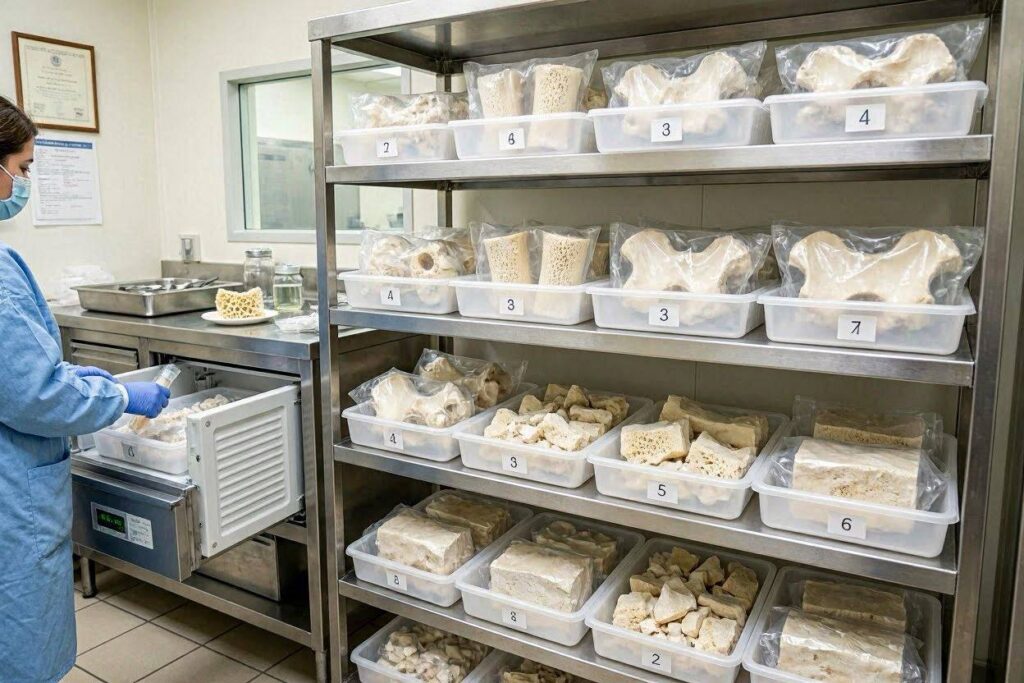
Once the recovered bone tissue arrives at the processing facility, it undergoes a sequence of treatments designed to remove all cellular material, including any potential pathogens, while preserving the bone’s mineral scaffold and collagen structure. This processing is the key reason why allograft bone does not require the recipient to take immunosuppressive drugs. Unlike a liver or heart transplant, a bone allograft is not a living tissue transplant. It is a structural matrix. The processing steps are proprietary but follow validated, FDA-cleared methods. These can include:
- A series of sterile water and alcohol baths to remove blood and lipid elements.
- Soaking in hydrogen peroxide or other agents to remove cellular debris.
- Freeze-drying (lyophilization) to remove water and create a shelf-stable product.
- A final, terminal sterilization step using gamma irradiation, a low-dose, controlled process that penetrates the tissue and breaks down the DNA/RNA of any potential residual pathogens without denaturing the bone morphogenetic proteins (BMPs), which are the natural growth factors that make the graft work.
The result is a sterile, biologically active but acellular scaffold that your body recognizes not as a foreign invader but as a familiar template for new bone growth.
| Safety Stage | Primary Action | Purpose |
|---|---|---|
| Donor Screening | In-depth medical/social history interview with next-of-kin; physical exam | Rule out donors with high-risk behaviors and diseases |
| Serological Testing | NAT and antibody tests on donor blood for HIV, HCV, HBV, Syphilis, HTLV | Directly detect viral genetic material, closing the window period |
| Aseptic Recovery | Surgical tissue removal in a sterile operating room by a trained team | Prevent introduction of environmental contaminants |
| Processing & Sterilization | Removal of cells/debris, freeze-drying, terminal gamma irradiation | Eliminate residual pathogens; inactivate viruses and bacteria; create an acellular matrix |
The Biological Role: Scaffolding for Your Own Cells
The term “cadaver bone” creates a false impression that a piece of dead person’s skeleton is simply screwed into your jaw. The biological reality is far more elegant and dynamic. The processed allograft bone is not a permanent implant in its original form. It is a temporary scaffold. Your body treats it as a blueprint for construction.
When a dentist places the allograft particles into your bone defect, they are filling a void with a three-dimensional, biologically compatible matrix. Your own blood soaks into this matrix, carrying with it circulating mesenchymal stem cells. These cells are the body’s master builders. They reach the allograft scaffold, adhere to its surface, and receive chemical signals—the surviving BMPs—that instruct them to transform into osteoblasts, the bone-forming cells. The osteoblasts then begin the slow, meticulous process of laying down a new, entirely your-own matrix of collagen and calcium phosphate. As they do this, they slowly resorb and replace the allograft scaffold through a process called creeping substitution. Over a period of months, the allograft material is completely removed by your body’s cells and replaced with living, vascular, native bone that is indistinguishable from the original jawbone. The donor tissue is gone, having served its purpose. What remains is a solid, living foundation of your own bone, ready to receive and permanently support a dental implant. You are not carrying around a piece of a deceased person. You have been healed by a process that used a human-derived matrix to guide your body to rebuild itself.
The Ethical and Emotional Terrain
The use of human donor tissue in any form treads on deeply personal, ethical, and emotional ground. Patients have a wide spectrum of reactions to the idea, all of which are completely legitimate. Some feel immense gratitude and are in awe of a system that allows such a gift. Others feel a visceral rejection or an association with death that they find profoundly uncomfortable.
From the perspective of the donor family, the act is almost universally framed as a search for meaning. In the aftermath of an unexpected death, a family is approached with a choice. They can leave the tragedy as a point of total destruction, or they can find within it a seed of life for others. The medical field is filled with anonymous recipients whose quality of life was restored because a grieving family said “yes” in their darkest hour. For a dental implant recipient, living with a smile rebuilt by such a gift can be a quiet, profound, and very personal reminder of human interconnectedness. For those who find the concept unacceptable, a clear alternative exists in the form of xenografts (bovine or porcine bone) or synthetic alloplast grafts, which use a man-made calcium phosphate matrix. An honest conversation with your surgeon about all available options is a crucial part of the pre-operative process.
The Regulatory Framework and the AATB
The safety and integrity of this entire system is not left to chance. In the United States, the Food and Drug Administration (FDA) regulates human cells, tissues, and cellular and tissue-based products (HCT/Ps) under Title 21, Part 1271 of the Code of Federal Regulations. This regulation mandates rigorous donor eligibility determination, current good tissue practice (cGTP), and comprehensive record-keeping and tracking from donor to recipient. This tracking means that if a problem were ever discovered with a donor, every piece of distributed tissue can be recalled.
Above and beyond the FDA’s baseline requirements, the accreditation by the American Association of Tissue Banks (AATB) is the gold standard in the industry. An AATB-accredited tissue bank has undergone voluntary, intensive on-site inspections and audits to verify that its processes meet or exceed the highest standards for donor screening, recovery, processing, storage, and distribution. When your dentist obtains an allograft product, they source it from a reputable manufacturer who uses AATB-accredited tissue banks. You can and should ask your dentist the specific name and manufacturer of the bone graft product they intend to use. A transparent surgeon will not hesitate to share this information and discuss why they trust that particular product.
Comparing Allograft to Other Graft Options
The choice of graft material is a clinical decision made by the surgeon based on the size and shape of the defect, the patient’s medical history, and the patient’s personal preferences. It is helpful to understand how cadaver bone (allograft) fits within the broader landscape of available materials.
- Autograft (Your Own Bone): Often considered the historical “gold standard” because it is 100% living, osteogenic tissue. It contains your own cells, growth factors, and a perfect collagen scaffold. The primary disadvantage is the need for a second surgical site, which carries its own morbidity, including pain, bleeding, and risk of nerve damage at the harvest site (typically the chin, back of the jaw, or hip). The volume of bone available is also limited.
- Allograft (Human Donor Bone): The subject of this guide. It provides a high-quality, osteoconductive scaffold without a second surgical site. It is acellular, so it is not osteogenic, meaning it does not provide living cells; it relies on your body’s cells to populate it. It is widely available in various forms.
- Xenograft (Animal Bone, usually Bovine): Processed to be completely deproteinized, leaving a pure mineral scaffold that is almost identical to human bone mineral. It resorbs extremely slowly, which is excellent for volume preservation over time. It is a very predictable material with a decades-long track record.
- Alloplast (Synthetic Material): These are entirely synthetic formulations of calcium phosphate, bioactive glass, or polymers. They have zero risk of biological contamination. They function purely as scaffolds and can be engineered to resorb at specific rates. They are a popular choice for patients who object to both human and animal-derived tissues.
The Practical Path from Donor to Dental Chair
Summarizing the logistical journey reveals the tightly controlled chain of custody. After the family consents and the recovery is complete, the donor is returned to the family for funeral arrangements with the utmost care, with no visible signs of the donation. The recovered tissue, labeled with a unique identification number, is sent on cold-chain transport to the processing facility. After weeks of processing and rigorous quality control testing, the final sterile product is freeze-dried into a powder, granules, or a moldable putty. It is packaged sterilely and sold to dental distributors, who then sell it to your dentist or oral surgeon. Your dentist stores it on a shelf in a dry, room-temperature environment until your scheduled grafting procedure. On the day of your surgery, the packet is opened in the sterile field, mixed with your own blood or sterile saline to form a workable graft material, and packed into your jaw. The donor’s journey, which ended in a recovery suite, begins again in the healing biology of your body.
Conclusion
Cadaver bone for dental implants, known clinically as human allograft, originates from the generous gift of registered donors whose tissue is recovered under strict sterile conditions. Its safety is ensured through an exhaustive, multi-layered system of medical history screening, advanced viral nucleic acid testing, and validated sterilization processes overseen by the FDA and accredited tissue banks. This processed tissue serves not as a dead implant but as a temporary biological scaffold that instructs your own body to resorb it and replace it with living, native bone, creating a solid foundation for a new tooth.
Frequently Asked Questions
Can I feel the “cadaver bone” inside my jaw forever?
No. You cannot feel the graft material, and over time, the allograft is entirely replaced by your own living bone through a process called creeping substitution. The final healed bone is biologically and structurally yours. There is no permanent piece of a donor in your jaw.
Is there a risk of my body rejecting the allograft like a heart transplant?
No. Rejection occurs when the immune system attacks living foreign cells. Processed allograft bone is acellular—all cells have been removed. It is a protein scaffold, not a living tissue. Therefore, the immune system does not see it as a threat, and no immunosuppressive drugs are needed.
Can I ask my dentist for a specific type of bone graft?
Absolutely. This is your body and your treatment. Discuss your concerns and ethical preferences openly. A good surgeon will explain their clinical reasoning for recommending one material but will also respect your values and explore alternatives like xenograft or alloplast if they are clinically appropriate for your specific case.
How does the dentist know the bone is from a safe donor?
Your dentist sources grafts from established, reputable dental supply companies. These companies are legally bound to provide products processed from tissue banks that follow FDA cGTP and ideally hold AATB accreditation. Your dentist can provide the product lot number and manufacturer, which links back to a fully documented and safe donor record.
Additional Resource:
To learn more about the standards of tissue banking, visit the official website of the American Association of Tissue Banks (AATB) at www.aatb.org.
Disclaimer: This article provides detailed information for educational purposes only. It is not intended as a substitute for professional medical or dental advice, diagnosis, or treatment. Always consult your dentist or oral surgeon regarding the specifics of your own medical condition and treatment plan.