What May Go Wrong with Dental Implants?
Dental implants boast a remarkable success rate, often cited above 95 percent. This statistic is a testament to decades of refined surgical techniques, advanced materials, and meticulous treatment planning. Yet, no medical procedure carries a zero-risk guarantee. The honest, responsible conversation about dental implants must include a clear-eyed look at what can go wrong. This comprehensive guide does not seek to alarm you but to empower you. Understanding the potential complications, their causes, their warning signs, and their treatments transforms you from a passive patient into an informed partner in your own care. We will explore both the early and late failures, the biological and mechanical problems, and the critical role of prevention.
Early Implant Failure: When Integration Does Not Occur
The biological success of a dental implant hinges on osseointegration, the direct structural and functional connection between living bone and the surface of the titanium implant. This process is a delicate biological dance. When it fails to occur, or when an initially successful integration breaks down before the implant is restored, it is termed early implant failure. This typically manifests within the first three to six months after placement. The patient may notice persistent discomfort, swelling, or, most tellingly, a loose implant that moves when wiggled. A healthy, integrating implant is rock-solid and asymptomatic.
The causes of early failure are multifactorial. A primary cause is surgical trauma. If the bone is overheated during the drilling process, the bone cells surrounding the implant site die, a condition called osteonecrosis. The implant is then sitting in dead bone, which cannot heal or integrate. Instead, the body walls off the implant with fibrous scar tissue, a clinical failure called fibro-osseous integration. Another surgical cause is a lack of primary stability. When the implant is placed, it must be firmly wedged into the bone with sufficient torque. If the bone quality is very soft, or if the surgeon inadvertently widens the preparation too much, the implant can micro-move during the healing phase. This micro-motion disrupts the delicate cellular processes of bone healing and encourages the formation of a fibrous capsule instead of a bony union.
Systemic and Patient-Related Risk Factors
Your general health plays a powerful role in the success of osseointegration. Uncontrolled systemic diseases severely impair healing. Patients with poorly controlled diabetes mellitus have compromised microcirculation and impaired white blood cell function, reducing the bone’s ability to heal and increasing the risk of post-operative infection. Autoimmune diseases and conditions requiring long-term corticosteroid therapy also depress the immune response and bone metabolism. Your surgeon must have a complete, honest picture of your medical history, including all medications and supplements you take, to accurately assess your individual risk profile.
Lifestyle choices are equally powerful determinants. Smoking is the single most significant modifiable risk factor for early implant failure. The vasoconstrictive effect of nicotine reduces blood flow to the gums and bone, starving the healing surgical site of oxygen and immune cells. Studies consistently show a two to three times higher failure rate in smokers compared to non-smokers. A history of intravenous bisphosphonate therapy, used to treat certain cancers and severe osteoporosis, is a critical red flag. These medications profoundly alter bone metabolism and can trigger medication-related osteonecrosis of the jaw, a devastating, non-healing condition of exposed bone, following even minor oral surgical trauma. Any patient with a history of bisphosphonate use must disclose this fully before any consideration of implant surgery.
Peri-Implantitis: The Silent, Progressive Threat
The most common cause of late implant failure, occurring after the implant has been successfully restored and in function for years, is peri-implantitis. This is a destructive inflammatory process affecting both the soft gum tissue and the hard bone supporting the implant. It begins innocuously as peri-implant mucositis, a reversible inflammation confined to the gum tissue. You may notice redness, swelling, and bleeding when you brush or floss around the implant. There is no bone loss at this stage, and it is completely curable with improved oral hygiene and a professional cleaning. The danger is that mucositis is often painless and easy to ignore.
If the bacterial biofilm that drives the inflammation is not eliminated, the inflammatory infiltrate extends deeper, breaching the weak biological seal where the gum attaches to the implant surface. The inflammation reaches the underlying bone, and the body begins to resorb it. This is now peri-implantitis. Bone loss around an implant is a unique and insidious process. Because the implant has no periodontal ligament and no nerve supply, significant bone destruction can occur silently, without pain, until the implant loses its support and becomes mobile. The pattern of bone loss is often a crater-shaped defect around the implant. The diagnosis is made by probing the gum pocket around the implant, noting bleeding and suppuration, and comparing current X-rays to baseline images to measure the loss of supporting bone height.
The Mechanisms of Bone Destruction
The disease process of peri-implantitis is more aggressive than periodontitis around a natural tooth. The absence of a periodontal ligament means there is no dense, well-vascularized barrier of collagen fibers to slow the apical migration of the bacterial front. Once the inflammatory process penetrates the weak mucosal seal, it spreads directly along the implant surface into the bone. The implant’s surface, which is intentionally roughened or treated to promote bone integration, becomes a liability. Once exposed to the oral environment by bone loss, this rough surface harbors a massive, complex biofilm that is incredibly difficult to decontaminate.
The body’s immune response, while intended to fight the bacteria, contributes to the destruction. Pro-inflammatory cytokines, released by immune cells in response to the bacterial challenge, stimulate osteoclast activity. Osteoclasts are the body’s bone-resorbing cells. They are sent into a state of overdrive, chewing away the bone that supports the implant. This is why treatment of peri-implantitis must not only target the bacteria but also modulate the host inflammatory response. The goal of treatment is to halt the disease progression, decontaminate the implant surface, and regenerate lost bone if possible. However, the bone loss is often irreversible, and the prognosis for an implant with advanced peri-implantitis is guarded.
Mechanical and Technical Complications
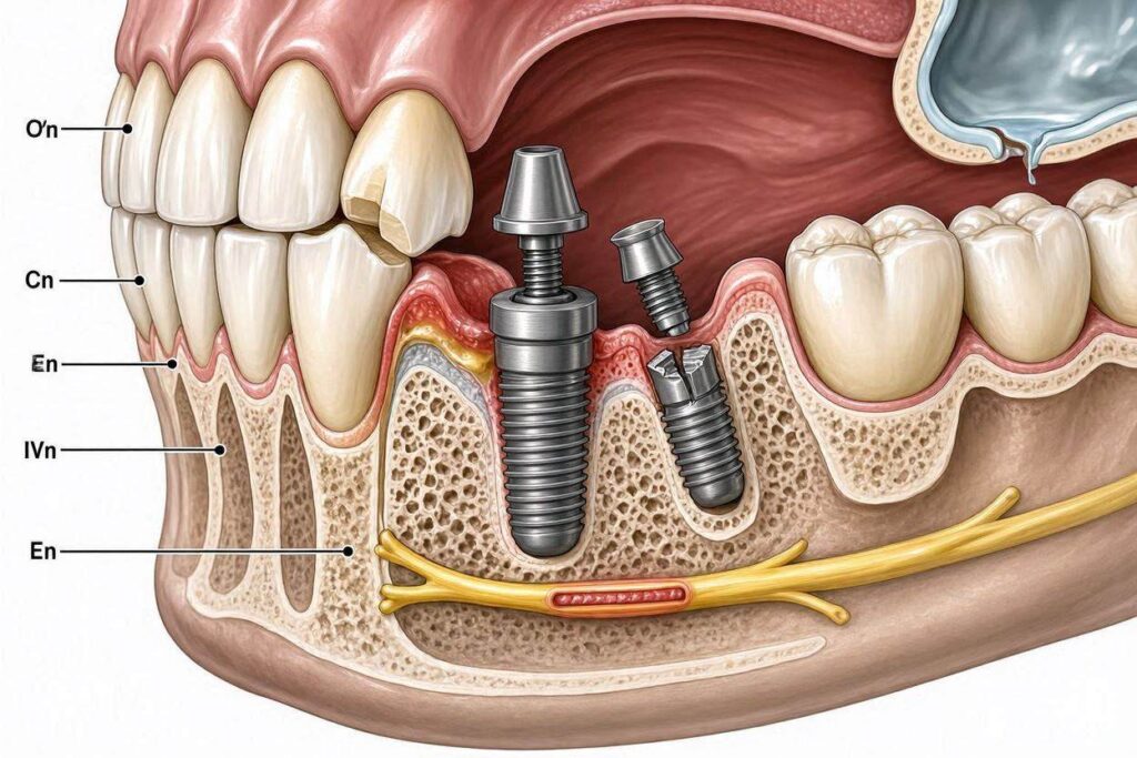
Not all implant problems are biological. The prosthetic components that attach to the implant and form the visible tooth are subject to mechanical forces and material fatigue. These complications are often less frightening than bone loss but are a significant source of inconvenience, additional expense, and treatment. The most common mechanical complication is the loosening of the abutment screw. This is the tiny titanium screw that connects the implant fixture in the bone to the abutment and the crown. Over years of chewing, the micro-movements can cause the screw to back out slightly, resulting in a feeling of looseness in the crown. Often, the crown itself is stable, but the internal connection has failed. This is usually a straightforward fix: the crown is removed, the screw is retorqued to the manufacturer’s specification, and the access hole is resealed.
A more serious mechanical failure is fracture of the implant fixture itself. This is rare with modern titanium alloy implants but can occur. Implant fracture is almost always a consequence of excessive, uncontrolled, off-axis forces over a long period. Patients with severe bruxism who do not wear a protective nightguard are at the highest risk. The constant lateral grinding forces create microfractures in the metal that eventually propagate into a complete break, often at the level of the first thread. A fractured implant is a catastrophic failure. The remnant embedded in the bone must be surgically removed, a complex and bone-destructive procedure, and after a healing period, a new implant may be placed, often in a slightly different position.
Veneering Material Fractures
The visible part of your implant restoration, the crown or the bridge, is made of a material designed to mimic the appearance of natural tooth enamel. This is most commonly porcelain fused to a metal or zirconia substructure, or a solid monolithic zirconia restoration. While these materials are strong, they are not indestructible. Chipping or fracturing of the veneering porcelain is a frustrating and relatively common complication. A small chip may be polished smooth. A larger fracture exposes the underlying substructure and requires a repair or a complete replacement of the prosthesis.
The causes of porcelain fracture are often a combination of material properties and occlusal forces. Some patients have a bite pattern that concentrates heavy forces on a single cusp of the implant crown, exceeding the cohesive strength of the porcelain. Again, bruxism is a major culprit. Improper design of the occlusion on the crown by the restorative dentist can also create damaging leverage and shear forces. The lesson is that mechanical complications are largely preventable through careful treatment planning, precise control of the biting forces, and the routine use of a custom-fitted occlusal nightguard for any patient with a history of clenching or grinding.
Surgical and Short-Term Post-Operative Complications
The surgical placement of dental implants is a remarkably safe procedure, but it involves a surgical wound in a complex anatomical area. Short-term complications, while typically manageable and transient, can occur. Pain, swelling, and bruising are normal expected consequences of the surgical trauma and usually peak within 48 to 72 hours. If pain suddenly intensifies after several days, rather than gradually improving, it raises the suspicion of an acute infection or a developing abscess. This is a medical urgency that requires immediate contact with your surgeon. A post-operative infection requires prompt antibiotic therapy and possibly surgical drainage to prevent it from compromising the integration of the implant.
Bleeding is expected for the first few hours, and persistent oozing can last a day or two. Significant, uncontrolled bleeding is rare and is often related to an undiagnosed bleeding disorder or the use of anticoagulant medications not properly managed pre-operatively. Another potential surgical complication is nerve injury. The mandibular nerve, which runs through the lower jaw and provides sensation to the lower lip and chin, is the structure of most concern. An implant placed too deeply or in the wrong position can compress, stretch, or transect this nerve. The result is paresthesia, an altered sensation ranging from numbness to tingling to a persistent burning pain in the lip and chin on the affected side. This can be temporary or, in the worst-case scenario, permanent. This devastating complication is almost entirely preventable with careful pre-operative 3D CBCT imaging and planning.
Sinus and Nasal Cavity Complications
When implants are placed in the upper back jaw, the maxillary sinus is the critical anatomical neighbor. The sinus is an air-filled cavity that often expands downward after tooth loss, leaving a thin floor of bone between the oral cavity and the sinus. If an implant is pushed too far superiorly, it can perforate the Schneiderian membrane that lines the sinus floor and protrude into the sinus cavity. This can cause a sinus communication, where air and fluids pass between the mouth and the sinus, leading to chronic sinus infections and sinusitis symptoms on that side.
A related complication is the displacement of an implant entirely into the sinus cavity. This is a surgical accident that requires an immediate referral to an oral surgeon or an otolaryngologist to retrieve the implant from the sinus, usually via a Caldwell-Luc procedure. Less dramatically, a sinus lift bone grafting procedure, often performed to increase bone height for implant placement, can be complicated by a tear of the delicate sinus membrane. If the tear is recognized during surgery, it is repaired. If it goes unrecognized, the graft material can migrate into the sinus, causing a chronic fungal or bacterial sinus infection. This highlights the absolute necessity of three-dimensional imaging and meticulous surgical technique in the posterior maxilla.
The Critical Role of Prevention and Early Intervention
The vast majority of the complications described in this guide are preventable. They are not random acts of misfortune. They are the predictable consequences of identifiable risk factors. The most powerful preventive tool in implant dentistry is a thorough, honest pre-operative assessment combined with advanced three-dimensional radiographic imaging. A surgeon who places an implant without a CBCT scan is navigating blindfolded. The scan reveals the exact three-dimensional topography of the bone, the precise location of nerves and sinuses, and any hidden pathology. It allows the surgeon to perform a virtual implant placement on a computer screen before ever touching the patient, selecting the ideal implant size and position to avoid all anatomical danger zones.
The second pillar of prevention is the meticulous optimization of patient health. An elective dental implant should be postponed in a patient with uncontrolled diabetes until their HbA1c level is within an acceptable range. A heavy smoker must be counseled that their implant carries a significantly higher risk of failure and that a smoking cessation program, even a temporary one during the healing phase, dramatically improves their prognosis. A patient with a history of periodontal disease must have that disease fully treated and stabilized before an implant is placed. Placing an implant in a mouth with active, untreated gum disease is like building a new house on a foundation riddled with termites. The underlying disease process will simply attack the new implant.
The Non-Negotiable Role of Professional Maintenance
After your implant is placed, restored, and functioning beautifully, the responsibility for prevention shifts to a partnership between you and your dental team. Your commitment to meticulous daily home care, as detailed in our guide on implant maintenance, is your frontline defense. The professional recall visit is your essential early warning system. You cannot skip these appointments. During a professional maintenance visit, the hygienist measures the gum pockets around your implant, checks for bleeding, and evaluates the occlusion. They take periodic X-rays, usually once a year, to compare with baseline images and detect any early, subtle bone loss.
This visit is also an opportunity for the dentist to evaluate the mechanical integrity of your restoration. They will check the tightness of the abutment screw, assess the occlusion for any new interferences, and inspect the porcelain for any hairline fractures or chips. They will ask you about any habits, like clenching or grinding, and ensure your nightguard fits properly. If early peri-implant mucositis is detected, a single intensive cleaning session can completely reverse it. If a loose screw is detected and tightened, a catastrophic fracture is prevented. Professional maintenance is not an optional service; it is the condition upon which your implant’s long-term warranty is written.
Conclusion
The vulnerabilities of dental implants span from early biological failure, driven by uncontrolled systemic disease, smoking, and surgical error, to the silent, progressive bone destruction of peri-implantitis fueled by bacterial biofilm. Mechanical complications, including screw loosening and porcelain fracture, arise primarily from excessive occlusal forces and bruxism, while serious surgical misadventures like nerve and sinus injury are almost entirely preventable with meticulous 3D imaging and planning. The ultimate safeguard against all these complications is a lifelong commitment to a tripartite partnership: optimized patient health, obsessive daily home care, and unwavering adherence to a schedule of professional maintenance and surveillance.
Frequently Asked Questions
What are the first signs of a failing dental implant?
The earliest warning signs are bleeding, redness, or swelling of the gum around the implant, a bad taste, or pus. A loose-feeling crown or a sensation of movement when chewing are serious signs that require immediate professional attention.
Can peri-implantitis be cured?
Peri-implant mucositis, the initial gum inflammation without bone loss, is completely reversible with treatment. True peri-implantitis, with bone loss, is a chronic condition that can be arrested and managed, but the lost bone is often irreversible. Early detection is critical.
Is it safe to get a dental implant if I smoke?
Smoking is a significant risk factor for both early and late implant failure due to impaired healing. While not an absolute contraindication, it dramatically increases your risk. A cessation program, especially during the surgical healing phase, is strongly recommended by all implantologists.
Why does my implant crown feel loose but nothing looks broken?
This is the classic sign of a loosened abutment screw. The internal screw connecting the crown to the implant fixture has likely backed out. This is a common mechanical complication that your dentist can often fix easily by retorquing the screw. Do not delay this repair.
Are implant complications more common in the upper or lower jaw?
The lower jaw presents the risk of nerve injury, while the upper back jaw presents the risk of sinus complications. The upper front aesthetic zone presents the challenge of managing thin gum tissue and potential aesthetic complications like gum recession. Each arch has its own specific risk profile.
Additional Resource
The International Congress of Oral Implantologists (ICOI) provides a wealth of scientific and patient-oriented information on implant dentistry, including the diagnosis and management of peri-implantitis. You can access their consensus papers and patient resources at: https://www.icoi.org/