Can Gum Tissue Cover Dental Implants?
You look in the mirror after your implant crown is placed, and something catches your eye. The gum around the implant looks different from the gum around your natural teeth. It may sit slightly lower, exposing a hint of metal. It may look thinner or paler. You wonder whether the gum tissue should cover the implant more completely, or whether what you are seeing is normal.
This question touches on one of the most important and least understood aspects of implant dentistry: the relationship between the soft tissue and the implant. The way gum tissue covers, surrounds, and seals around an implant affects not only the aesthetics of your smile but the long-term health and survival of the implant itself. This guide explains everything you need to know about gum tissue and dental implants. You will learn what is normal, what is problematic, what can be done to improve soft tissue coverage, and how to maintain healthy gums around your implants for a lifetime.
The Anatomy of Gum Tissue Around Natural Teeth and Implants
To understand how gum tissue interacts with implants, you first need to understand how it interacts with natural teeth. The differences between these two relationships explain most of what you observe around your implant.
The Natural Tooth-Gum Interface
Around a natural tooth, the gum tissue forms a specialized attachment. A cuff of keratinized gingiva, the firm, pink gum tissue that is resistant to chewing forces, encircles the tooth. Below the visible gum line, the gum tissue attaches to the tooth root via a structure called the junctional epithelium. Deeper still, connective tissue fibers called Sharpey’s fibers insert directly into the cementum covering the root surface. These fibers act like tiny guy wires, anchoring the gum tightly to the tooth.
This biological attachment creates a seal that prevents bacteria from penetrating into the underlying bone. It also provides a resilient, aesthetically pleasing frame for the tooth. The gum tissue between the teeth, the interdental papilla, fills the triangular space beneath the contact point, creating a natural scalloped contour.
The Implant-Gum Interface
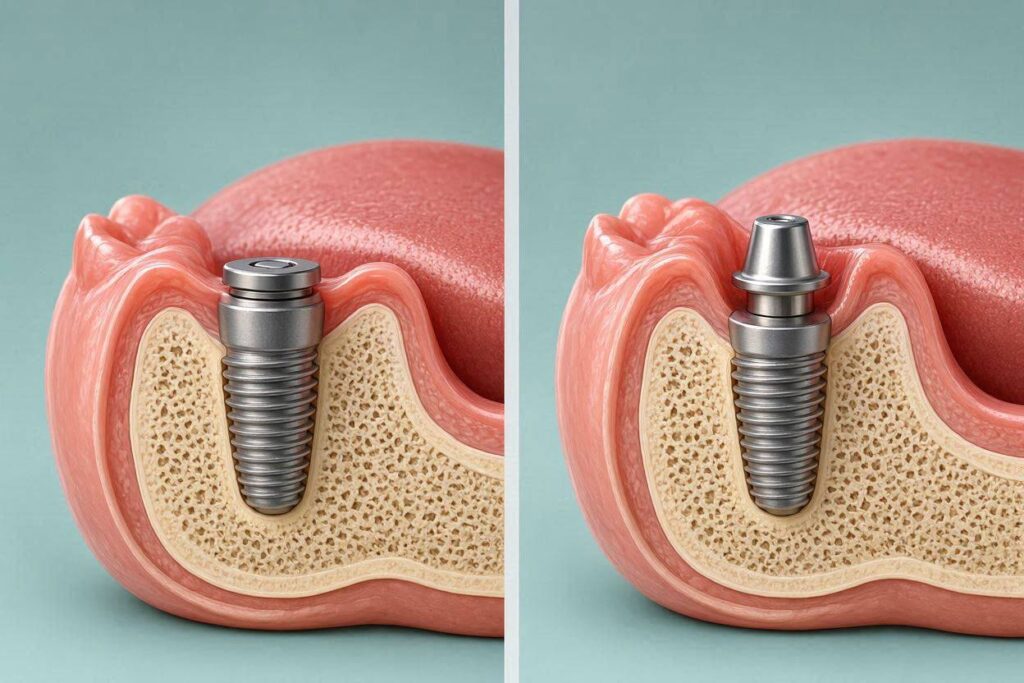
Around a dental implant, the attachment is fundamentally different. The implant surface is titanium or zirconia, not cementum. The junctional epithelium attaches to the implant surface, but the connective tissue fibers cannot insert into the implant. Instead, they run parallel to the implant surface or form a circular cuff around it. The tight mechanical interlock that exists around a natural tooth does not exist around an implant.
This difference has practical consequences. The seal around an implant is less robust than the seal around a natural tooth. Bacteria can penetrate more easily if oral hygiene is inadequate. The gum tissue around an implant is also more vulnerable to recession and may not fill the interdental spaces as predictably as gum tissue around natural teeth.
What Normal Gum Coverage Looks Like Around an Implant
The first step in addressing concerns about gum coverage is understanding what a healthy, normal implant site looks like.
The Transmucosal Zone
The implant abutment, the connector between the implant body and the crown, passes through the gum tissue. This is the transmucosal zone. The gum forms a cuff around the abutment, creating a collar of soft tissue that is 2 to 4 millimeters thick. This cuff should look pink and firm, without redness, swelling, or bleeding when probed gently.
The crown should emerge from this cuff naturally, without visible metal. The transition from the round abutment to the anatomically shaped crown should occur at or slightly below the gum line. In a well-designed implant restoration, you see the crown emerging from the gum, just as you see a natural tooth emerging. You do not see the abutment or the implant body.
The Interdental Papilla
The small triangle of gum tissue that fills the space between the implant crown and the adjacent tooth is the interdental papilla. The presence or absence of this papilla makes a dramatic difference in the appearance of the implant.
In natural teeth, the papilla fills the space completely when the distance from the contact point between the teeth to the bone crest is 5 millimeters or less. Around implants, this distance is even more critical. The papilla around an implant is supported primarily by the bone on the adjacent natural tooth, not by the implant itself. If the adjacent tooth has healthy bone, the papilla may be present. If the adjacent tooth has lost bone, or if the implant is next to another implant, the papilla may be missing, creating a black triangle between the teeth.
What You Should and Should Not See
In a successful implant restoration, you should see a natural-looking crown emerging from healthy pink gum tissue. You should not see exposed implant threads, which are the roughened surface of the implant body designed to be in contact with bone. You should not see a gray shadow of metal showing through the gum, though this can occur in patients with thin gum tissue. You should not see persistent redness, swelling, or bleeding.
When Gum Tissue Does Not Cover the Implant Properly
Several situations can result in inadequate or abnormal gum coverage around an implant. Understanding the cause is essential to finding the right solution.
Implant Thread Exposure
The roughened surface of an implant is designed to be buried in bone. When this surface becomes exposed to the oral environment, it is a problem. Bacteria colonize the rough surface, and the patient cannot clean it effectively. The exposure may result from gum recession, inadequate bone covering the implant at placement, or surgical placement of the implant in a position where the bone was too thin to cover it fully.
Implant thread exposure is a risk factor for peri-implantitis, the inflammatory condition that causes bone loss around implants. If you can see or feel the rough surface of your implant, you need to see your dentist. The situation may require surgical intervention to cover the exposed surface.
Gum Recession Around Implants
Gum tissue can recede around implants just as it can around natural teeth. Recession exposes the abutment or the implant surface and creates an aesthetic concern, particularly in the front of the mouth. Recession occurs for several reasons.
The implant may have been placed too far toward the cheek or lip, leaving insufficient bone and gum tissue on the facial side. The gum tissue may be thin and fragile to begin with. Aggressive tooth brushing can traumatize the gum and cause it to recede. Orthodontic movement or occlusal forces can contribute. Inflammation from inadequate oral hygiene can cause the gum to pull away.
Once gum recession occurs around an implant, it rarely reverses without surgical intervention. The focus should be on identifying the cause and stabilizing the situation before the recession worsens.
Inadequate Keratinized Tissue
Keratinized gingiva is the firm, resilient gum tissue that is tightly bound to the underlying bone. It is the type of gum that resists the mechanical forces of chewing and tooth brushing. Around some implants, the band of keratinized tissue is very narrow or absent entirely. Instead, the implant is surrounded by alveolar mucosa, the looser, darker, more movable tissue that lines the cheeks and the floor of the mouth.
A lack of keratinized tissue around an implant makes oral hygiene more difficult. The movable tissue is harder to clean, more prone to inflammation, and less resistant to recession. Patients with inadequate keratinized tissue may benefit from a soft tissue graft to create a band of firm, attached gingiva around the implant.
Surgical Procedures to Improve Gum Coverage
Modern periodontics and implant dentistry offer several procedures to improve the quantity and quality of gum tissue around implants. These procedures fall under the umbrella of soft tissue management and are typically performed by a periodontist or an implant surgeon with advanced training.
Connective Tissue Grafts
A connective tissue graft is the most common procedure for increasing the thickness and coverage of gum tissue around an implant. A small piece of connective tissue is harvested from the roof of the patient’s mouth, from an area behind the back teeth. The donor site heals quickly, similar to a minor burn on the palate.
The harvested tissue is placed under the gum around the implant, increasing the bulk and thickness of the soft tissue. The graft also brings blood supply to the area, which improves the health and resilience of the overlying gum. Over time, the graft integrates with the existing tissue, creating a thicker, more stable soft tissue cuff around the implant.
Connective tissue grafts can be performed at the time of implant placement, at the time of abutment connection, or after the implant has been restored if recession or thin tissue becomes apparent. The earlier the graft is placed, the better the aesthetic outcome tends to be, because the graft can shape the tissue before the final crown is fabricated.
Free Gingival Grafts
A free gingival graft is used when the primary goal is to increase the width of keratinized tissue around an implant. Unlike a connective tissue graft, which is placed under the existing gum, a free gingival graft includes the surface epithelium and is placed on top of the prepared recipient site.
The graft is harvested from the palate, usually from an area farther forward than the connective tissue graft donor site. The recipient site is prepared by removing a strip of the non-keratinized alveolar mucosa, creating a bed for the graft. The graft is sutured into place and heals over several weeks. The result is a band of firm, keratinized tissue that resists recession and facilitates oral hygiene.
Free gingival grafts are less aesthetic than connective tissue grafts because the color and texture of the graft tissue may not match the surrounding gum perfectly. For this reason, they are typically used in the back of the mouth where aesthetics are less critical.
Pinhole Surgical Technique and Tunneling Procedures
Minimally invasive techniques have been developed to reposition gum tissue without harvesting a graft from the palate. The pinhole surgical technique involves making a small hole in the gum and using specialized instruments to loosen and reposition the tissue over the exposed implant or tooth root. Tunneling procedures create a pouch under the gum into which graft material can be inserted without large incisions.
These techniques are less invasive, have less postoperative discomfort, and heal faster than traditional grafting procedures. They are suitable for mild to moderate recession cases. Severe recession with significant tissue deficiency may still require a traditional graft.
The Timing of Soft Tissue Procedures
When a soft tissue graft is performed in relation to implant placement affects both the outcome and the overall treatment timeline.
Grafting at Implant Placement
Placing a connective tissue graft at the same time as the implant fixture offers several advantages. The graft can increase the thickness of the tissue overlying the implant from the very beginning of the healing period. This thick tissue protects the underlying bone graft if one was placed and creates a better emergence profile for the eventual crown.
The disadvantage is that grafting adds time and complexity to the implant surgery. The patient must manage both the implant surgical site and the palate donor site simultaneously. Some swelling and discomfort are expected.
Grafting at Abutment Connection
When a two-stage implant protocol is used, the implant is buried under the gum for the healing period. At the second-stage surgery, when the implant is exposed and a healing abutment is placed, a connective tissue graft can be performed. This timing allows the surgeon to place the graft in the exact area where it is needed most, guided by the position of the implant.
Grafting at second-stage surgery also allows any bone grafting placed at the first surgery to heal before the soft tissue procedure. The patient undergoes two surgeries, but each is shorter and less complex than a combined procedure.
Grafting After Restoration
Soft tissue grafting can be performed after the implant crown has been in place for some time if recession or thin tissue becomes a problem. This is a salvage procedure, addressing a complication that has developed. The results may not be as aesthetic as grafting performed earlier in the process, but significant improvement is possible.
Post-restoration grafting is more technically demanding because the crown and abutment must be removed or worked around. The tissue contours that have been established by the crown are disrupted and must be reestablished. Whenever possible, soft tissue deficiencies should be addressed before the final crown is fabricated.
Non-Surgical Management of Gum Health Around Implants
Not every concern about gum coverage requires surgery. Excellent daily care and professional maintenance can keep the gum tissue around implants healthy and stable.
The Importance of Daily Oral Hygiene
Implants require the same meticulous daily care as natural teeth, and in some ways, more. The gum attachment around an implant is less resistant to bacterial penetration, so plaque control is critical.
Brush around the implant at least twice daily with a soft-bristled toothbrush. Pay particular attention to the area where the crown meets the gum. Use an interdental brush, floss designed for implants, or a water flosser to clean between the implant and the adjacent teeth. A water flosser with a non-metal tip is especially useful for cleaning under implant-supported bridges and around full-arch prostheses.
If your implant restoration has areas you cannot reach with a brush or floss, talk to your dentist or hygienist. They can recommend specialized cleaning aids and demonstrate the proper technique.
Professional Maintenance Visits
You should see your dentist or hygienist for professional maintenance at least twice a year, and possibly every three to four months if you have risk factors for peri-implant disease. During these visits, the clinician will check for signs of inflammation, measure probing depths around the implant, take X-rays periodically to monitor bone levels, and clean the implant surfaces with specialized instruments.
The instruments used to clean implants are different from those used on natural teeth. Titanium and carbon fiber scalers clean the implant and abutment surfaces without scratching them. Scratches on an implant surface create niches for bacteria and are difficult to polish smooth. Your hygienist should be trained in implant maintenance and use instruments appropriate for the type of implant you have.
Managing Risk Factors
Certain factors increase the risk of gum problems around implants. Smoking is the single most significant modifiable risk factor. Smokers have higher rates of implant failure and peri-implantitis. If you smoke, quitting or reducing smoking dramatically improves the long-term health of the gum tissue around your implants.
Poorly controlled diabetes also increases risk. Work with your physician to manage your blood sugar. Inflammatory conditions and medications that cause dry mouth can affect peri-implant health. Tell your dentist about all medical conditions and medications so that your maintenance schedule can be adjusted accordingly.
The Aesthetic Aspect of Gum Coverage
For implants in the front of the mouth, the appearance of the gum tissue is as important as the appearance of the crown. A beautiful crown emerging from unhealthy or receded gum tissue is not a beautiful result.
The Pink Aesthetic Score
Dental researchers use a scale called the pink aesthetic score to evaluate the appearance of the soft tissue around implant restorations. The score assesses the presence and shape of the interdental papilla, the level and contour of the gum margin, the color and texture of the gum tissue, and the overall harmony with the adjacent natural teeth.
A high pink aesthetic score indicates that the soft tissue looks natural and healthy. Achieving this outcome requires careful planning, precise surgical technique, and, often, soft tissue grafting. Patients who place a high value on the appearance of their smile should discuss the pink aesthetic with their implant team before treatment begins.
The Role of the Provisional Restoration
A provisional restoration, a temporary crown placed on the implant during the healing period, can be used to sculpt the gum tissue. The provisional crown is shaped to support the gum in the desired position and contour. Over several weeks or months, the gum adapts to the provisional, creating a natural emergence profile.
When the final crown is fabricated, the laboratory technician replicates the contours of the provisional that created the ideal soft tissue architecture. This technique, called progressive loading or soft tissue conditioning, is a standard approach for achieving optimal aesthetics in the anterior zone.
Conclusion
Gum tissue can and should cover the implant abutment and the top of the implant body, creating a healthy pink cuff through which the crown emerges naturally. The gum attachment around an implant is structurally different from that around a natural tooth, lacking the connective tissue fiber insertion that creates a tight seal. This makes implants more vulnerable to recession and bacterial penetration. When gum coverage is inadequate, surgical procedures such as connective tissue grafts, free gingival grafts, and minimally invasive tunneling techniques can improve soft tissue quantity and quality. Meticulous daily hygiene, professional maintenance, and management of risk factors are essential to preserving gum health around implants for a lifetime.
Frequently Asked Questions
Can gums grow back around an implant after recession?
Gum tissue does not spontaneously regenerate around implants once recession has occurred. Surgical grafting is usually required to restore lost tissue. Preventing recession through proper implant placement, good oral hygiene, and avoidance of aggressive brushing is the best strategy.
Is it normal to see a little metal at the gum line of my implant?
A very small margin of the abutment may be visible if your gum tissue is thin, but ideally, you should not see metal. If the metal is visible and bothers you, a soft tissue graft or a new crown with a different margin design may improve the appearance.
How do I know if my gums around my implant are healthy?
Healthy peri-implant gums are pink, firm, and do not bleed when you brush or floss. There should be no swelling, redness, pus, or bad taste. Regular professional examinations are essential to detect problems before they become visible to you.
Can I have gum graft surgery if I have multiple implants?
Yes, soft tissue grafting can be performed around multiple implants. The procedures may be staged to manage patient comfort, addressing one area at a time or combining sites in the same quadrant.
Does gum grafting around an implant hurt?
The procedure is performed under local anesthesia and should not be painful. Post-operative discomfort is usually mild to moderate and managed with over-the-counter pain relievers. The donor site on the palate may feel like a minor burn for a few days and heals within one to two weeks.
Additional Resource:
For information on peri-implant soft tissue management, visit the American Academy of Periodontology at https://www.perio.org.