How Dental Implants Fuse to Bone?
Losing a tooth changes more than your smile. It affects how you chew, speak, and feel about yourself. Dental implants offer a remarkable solution. They look, feel, and function like natural teeth. But what makes them so stable? The answer lies in a fascinating biological process. The implant doesn’t just sit in your jaw. It becomes part of it. This article explores exactly how dental implants fuse to bone. We will break down the science, the stages, and the factors that influence success. You’ll understand why this process is the cornerstone of modern implant dentistry.
Understanding Osseointegration
The fusion of a dental implant to bone has a scientific name. It’s called osseointegration. This term comes from the Greek word “osteon” for bone and the Latin word “integrare” for making whole. A Swedish orthopedic surgeon, Per-Ingvar Brånemark, discovered this phenomenon by accident in the 1950s. He placed titanium chambers in rabbit bones for a study. When he tried to remove them later, he found the bone had adhered so tightly to the titanium that they couldn’t be separated. This happy accident revolutionized dentistry.
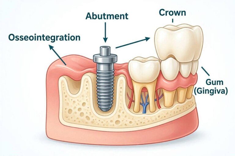
Osseointegration refers to the direct structural and functional connection between living bone and the surface of a load-bearing implant. In simpler terms, your jawbone cells attach directly to the implant’s surface. There is no soft tissue, cartilage, or ligament fibers between them. This is different from a natural tooth, which has a periodontal ligament acting as a shock absorber. An implant doesn’t have this ligament. It connects rigidly to the bone. This direct anchorage provides incredible stability. It’s what allows a small titanium post to support the tremendous forces of chewing day after day.
The Biological Significance
Why is this direct contact so crucial? When bone grows onto the implant surface, it locks the fixture in place mechanically. This prevents micro-movements that could cause the implant to fail. The intimate contact also creates a seal. This seal prevents bacteria and oral fluids from migrating down alongside the implant. The bone cells essentially recognize the implant surface as a suitable structure to attach to. They remodel and adapt continuously, just like bone elsewhere in the body. This living connection means the implant can handle stress. Under load, the bone around the implant strengthens. Without load, it can shrink. This dynamic relationship keeps the implant stable for decades.
The Science Behind Bone-Implant Fusion
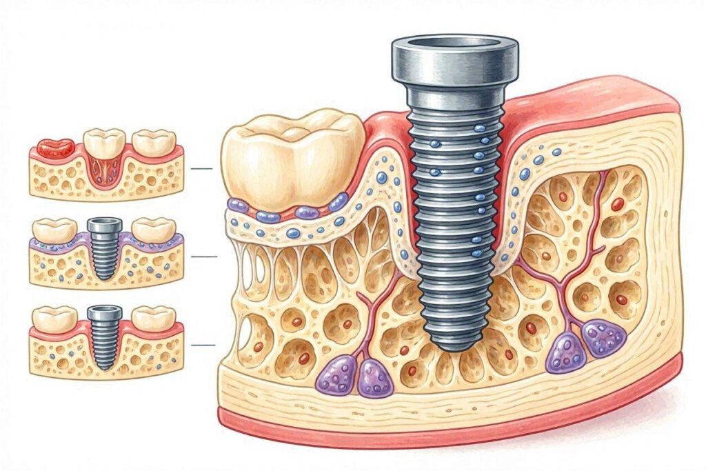
At a microscopic level, osseointegration is a marvel of biology. The implant surface, usually made of commercially pure titanium or a titanium alloy, has a special property. It forms a titanium oxide layer spontaneously when exposed to air. This oxide layer is highly biocompatible. It allows proteins from your blood to adsorb onto the surface immediately after the implant is placed. These proteins trigger a cascade of events. They attract bone-forming cells called osteoblasts. The osteoblasts migrate to the implant surface and begin laying down a matrix of collagen and other proteins. This matrix then mineralizes. Calcium phosphate crystals deposit within it, creating new bone directly on the titanium oxide layer.
The Role of the Titanium Oxide Layer
Think of the titanium oxide layer as a biological welcome mat. It is only a few nanometers thick, but it is incredibly stable. It resists corrosion from bodily fluids. More importantly, its surface chemistry and topography encourage bone cell adhesion. Modern implants have surfaces that are deliberately roughened at the microscopic level. Techniques like sandblasting and acid etching create peaks and valleys. This increases the surface area dramatically. Osteoblasts love this rough surface. They anchor themselves to it more effectively than to a smooth surface. The bone grows into these microscopic irregularities, creating a powerful interlocking bond. This is not just a chemical bond but also a mechanical interlock at the cellular level.
Cellular Events During Osseointegration
The process begins with a blood clot forming around the implant. This clot is rich in platelets and growth factors. These signaling molecules call in undifferentiated mesenchymal cells. These cells can transform into different types of connective tissue cells. In the presence of the implant and with proper stability, they become osteoblasts. The osteoblasts first lay down a layer of non-calcified bone matrix called osteoid. This osteoid then mineralizes. This initial bone is called woven bone. It forms quickly but is not very organized. Over the following months, this woven bone is remodeled. It is replaced by mature, lamellar bone. This lamellar bone is highly organized and strong. It is packed with blood vessels that sustain the living tissue around the implant.
The Osseointegration Process: A Timeline
Osseointegration is not an instant event. It is a gradual process that unfolds over several months. The timeline can vary based on the patient’s health, bone quality, and the implant location. However, a general pattern exists. Understanding this timeline helps manage expectations. It explains why you must wait for a crown after implant placement.
Initial Healing Phase: Days 1-7
Immediately after the implant is placed into the jawbone, the body’s healing response kicks in. The surgical site is essentially a controlled wound. A blood clot forms in the tight space between the implant threads and the surrounding bone. This clot is vital. It acts as a temporary scaffold. Platelets within the clot release growth factors like platelet-derived growth factor (PDGF) and transforming growth factor-beta (TGF-β). These signal immune cells to clean the area. They also attract cells that will build new tissue. During this first week, the goal is to protect the clot. This is why dentists give strict instructions. You must avoid disturbing the surgical area. Sucking through a straw, vigorous rinsing, or touching the site can dislodge the clot. Losing the clot severely compromises osseointegration.
Bone Formation Phase: Weeks 2-6
Around the second week, the real bone-building begins. New blood vessels form, a process called angiogenesis. They bring oxygen and nutrients to the healing site. The undifferentiated mesenchymal cells, guided by growth factors, differentiate into osteoblasts. These bone-forming cells start secreting osteoid onto the implant surface. This happens first where the implant is in closest contact with the old bone, such as the tips of the threads. This woven bone is deposited rapidly. By week four, the implant gains some initial stability from this new bone. However, this woven bone is weak. It has a random fiber orientation, like plywood made with chaotic layers. The implant cannot yet withstand full chewing forces. Premature loading at this stage can cause the implant to wobble. This micro-motion encourages the formation of fibrous scar tissue instead of bone. This is a leading cause of early implant failure.
Bone Remodeling Phase: Months 2-6 and Beyond
From about two months onward, the body starts a remodeling process that can continue for life. The weak, disorganized woven bone is gradually resorbed by cells called osteoclasts. In its place, osteoblasts lay down new, organized lamellar bone. This bone has layers of collagen fibers aligned in parallel, making it far stronger. The bone remodels along lines of stress. This is Wolff’s Law in action: bone adapts to the loads placed upon it. When the implant is eventually loaded with a crown, the forces of chewing are transmitted to the surrounding bone. This stimulates the bone to maintain its density and even strengthen around the implant. The interface between the implant and the bone becomes a dynamic, living connection. Even after full integration, the bone around a healthy implant typically renews itself at a rate of about 0.5-1% per year.
Key Factors Influencing Successful Bone Fusion
Not everyone heals the same way. Several factors play a critical role in determining whether an implant successfully fuses to bone. Dentists carefully evaluate these factors during the treatment planning phase. Recognizing them helps you understand your own risk profile.
Biocompatibility of Implant Material
The material of the implant is the foundational factor. Titanium and its alloys remain the gold standard due to their exceptional biocompatibility. The spontaneously forming oxide layer is the secret. Zirconia is a metal-free, ceramic alternative that also exhibits excellent osseointegration properties. Zirconia implants are relatively newer but have shown bone-to-implant contact rates similar to titanium in studies. The body does not reject these materials in the way it rejects transplanted organs. There is no immune reaction against the material itself. Instead, the body treats it as a scaffold for new bone.
Implant Surface Topography and Chemistry
As mentioned, a smooth surface is less effective for bone fusion than a rough one. Modern implants undergo surface modifications to increase their “osteoconductivity.” This means they encourage bone to grow along their surface. Common surface treatments include:
- Sandblasting with large grit and acid etching (SLA): This creates macro and micro-roughness.
- Anodization: An electrochemical process that thickens the oxide layer and creates a porous surface.
- Hydroxyapatite coating: A calcium phosphate compound that chemically mimics natural bone mineral, actively stimulating bone formation.
The difference can be significant. A moderately roughened surface can show 50% to 100% higher removal torque values (a measure of osseointegration strength) compared to a machined smooth surface.
Primary Stability
Primary stability is the mechanical engagement of the implant with the bone immediately after placement. It’s like a screw’s initial tight grip in wood. This is essential for osseointegration. If the implant moves, even microscopically, osteoblasts cannot form a direct bone connection. Instead, fibroblasts will seal the implant with a fibrous capsule, like scar tissue. This is a failed integration. A dentist achieves primary stability by using precise drilling protocols and tapping the implant into a site slightly smaller than its diameter. Bone density and volume are crucial here. Dense, thick bone in the lower front jaw provides better primary stability than the soft, spongy bone often found in the upper back jaw.
Bone Quality and Quantity
The jawbone is not the same everywhere. The best bone for an implant is dense, thick, and well-vascularized. Bone is classified into types:
- Type I (Dense Cortical): Excellent for stability but has fewer blood vessels, so healing can be slower.
- Type II (Thick Cortical, Dense Cancellous): Ideal for implants.
- Type III (Thin Cortical, Dense Cancellous): Most common in the upper jaw, still good for implants.
- Type IV (Very Thin Cortical, Fine Cancellous): Found in the posterior upper jaw. Provides poor initial stability and osseointegration predictability.
If a tooth has been missing for a long time, the bone in that area shrinks. This is called resorption. Without a tooth root stimulating the bone, the body thinks the bone is no longer needed. A cone-beam CT scan allows the dentist to assess the available bone in three dimensions. If there is not enough bone, a bone graft procedure may be necessary before or during implant placement.
Surgical Technique and Temperature Control
A skillful, atraumatic surgical technique is non-negotiable. The drills used to prepare the implant site generate heat due to friction. Excessive heat is a death sentence for bone cells. If the bone temperature exceeds 47°C (116.6°F) for more than one minute, the bone tissue can undergo necrosis. It essentially dies. The dead bone will not osseointegrate. Instead, it will be resorbed by the body, and fibrous tissue will replace it. To prevent this, the surgeon uses copious sterile saline irrigation to cool the drill. They use sharp, new drills and an intermittent drilling technique, lifting the drill periodically. Gentle handling of the soft tissue and bone preserves the blood supply, which is critical for healing.
Patient-Related Factors
Your overall health and habits directly impact bone healing.
- Smoking: This is one of the most significant risk factors for implant failure. Nicotine causes vasoconstriction, shrinking the small blood vessels that bring healing cells and oxygen. Smokers can have an implant failure rate two to three times higher than non-smokers.
- Uncontrolled Diabetes: High blood sugar impairs white blood cell function and wound healing. Patients with poorly controlled HbA1c levels have a much higher risk of peri-implant complications.
- Osteoporosis: This condition itself is not a strict contraindication, but certain medications for it, particularly oral bisphosphonates, have been associated with a rare but serious risk of medication-related osteonecrosis of the jaw (MRONJ).
- Oral Hygiene: A clean mouth is mandatory. Plaque buildup around a healing implant can cause inflammation in the surrounding gum, which can spread to the forming bone-implant interface.
Signs and Symptoms of a Failing Osseointegration
A successfully integrating implant should feel like nothing at all. The process is quiet and painless. However, a problem can arise. Being aware of warning signs is essential for early intervention.
What to Watch For During Healing
In the weeks and months after surgery, you must be vigilant. A healing implant site should have healthy, pink, and firm gum tissue. There should be no persistent swelling, no pus, and no throbbing pain that isn’t relieved by initial post-operative medications. A key warning sign is mobility. A healing implant should have absolutely no visible or palpable movement. If you can feel a wiggle when you gently touch it with your tongue, or if your dentist notes movement during a check-up, it’s a major red flag. Another sign is a dull ache or pain when you tap on the implant, indicating that the interface is not solid.
The Difference Between Soft Tissue and Hard Tissue Failure
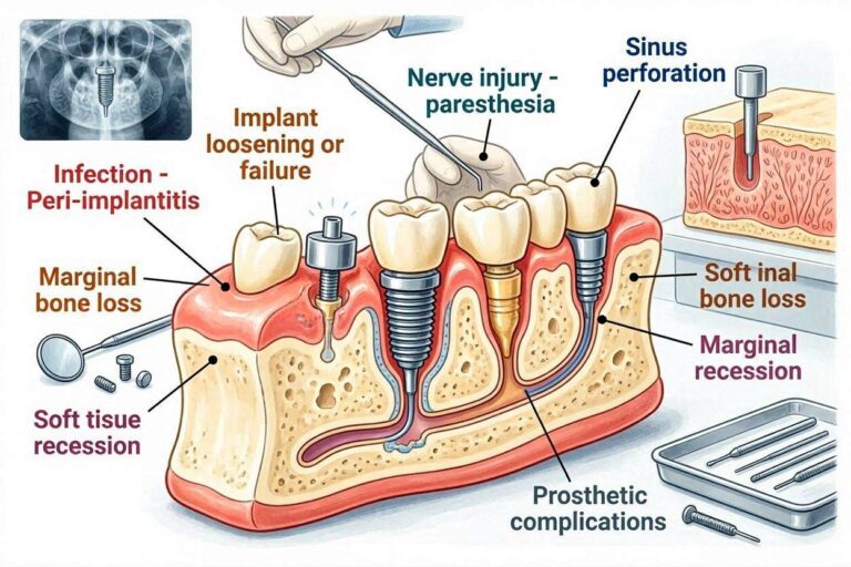
Early failure is often a failure of osseointegration. The bone and implant fail to fuse. Instead, a layer of fibrous tissue encapsulates the implant. This can result from overheating the bone, premature loading, lack of primary stability, or poor bone quality. The implant will feel loose. There might be radiolucency (a dark line) around the implant on an X-ray. A late failure occurs after the implant has functioned successfully for some time, often years. This is usually due to peri-implantitis, a bacterial infection that destroys the supporting bone. The implant might still feel integrated, but an X-ray shows crater-like bone loss around it. The gums will be red, swollen, and bleed easily. Treating peri-implantitis is complex, making prevention through excellent oral hygiene the best strategy.
How Dentists Confirm That the Implant Has Fused
Dentists don’t just guess when the implant is ready. They use objective and subjective methods to assess the level of osseointegration before placing the final tooth. Jumping to the restorative phase too soon can overload the immature bone-implant interface and ruin months of healing.
Clinical Examination Methods
The simplest test is the percussion test. The dentist taps the implant with a metal instrument. A successfully integrated implant emits a clear, high-pitched, crystalline ring. A failing, fibrous-encapsulated implant yields a dull, low-pitched thud. The absence of clinical mobility is the single most important sign. The dentist may use two instrument handles to try to rock the implant from side to side. Any movement greater than a minuscule flex within the bone is unacceptable. The condition of the surrounding peri-implant soft tissue is also checked. It must be firm, keratinized, and free of inflammation. Bleeding on gentle probing is a sign of peri-implant mucositis, which is a risk.
Imaging and Instrumentation
X-rays are a critical tool. A baseline X-ray is taken immediately after surgery. Periodic X-rays are then compared to this baseline. A healthy osseointegrated implant will show intimate contact between the bone and the implant surface with no intervening dark line. Bone levels should be maintained at the top of the implant or the first thread. Any radiolucency around the entire implant body is a sign of complete failure.
More advanced methods provide quantitative data. Resonance Frequency Analysis (RFA) is a non-invasive technique. A small magnetic peg is attached to the implant. A device emits a magnetic pulse, making the peg vibrate. The more stable the implant in the bone, the higher the resonance frequency. This value is displayed as an Implant Stability Quotient (ISQ) on a scale of 1 to 100. A stable, integrated implant typically has an ISQ over 65-70. A rising ISQ value over time indicates progressive osseointegration. The reverse torque test, where a dentist applies a specific torque in the opposite direction, is less favored. It risks damaging a healing interface. If an implant can withstand a removal torque of 35 Ncm without moving, it is generally considered ready for loading.
Enhancing Osseointegration: Modern Advances
The journey to faster and more reliable bone fusion is a hot topic in dental research. Significant advances are changing patient experiences.
Surface Technology Breakthroughs
Scientists are engineering implant surfaces that are not just rough but also bioactive. Hydrophilic surfaces are a leap forward. Traditional SLA surfaces are hydrophobic, repelling water and slowing the initial protein adsorption. New chemical treatments create a surface that attracts water and, consequently, proteins and cells. This dramatically speeds up the first hours of healing. Clinical studies show these hydrophilic implants achieve high secondary stability much faster, often allowing for early or immediate loading protocols in select cases. Another frontier is the use of nanotechnology. Creating surface features at the nanoscale (billionths of a meter) directly influences cell behavior. These structures can mimic the natural nanotexture of bone collagen, guiding osteoblast adhesion and differentiation with incredible precision.
Biological and Systemic Modulators
The use of growth factors is a powerful strategy. One example is recombinant human bone morphogenetic protein-2 (rhBMP-2). This protein is a potent osteoinductive signal. It can actually stimulate the patient’s own stem cells to differentiate into bone-forming cells. It has been used successfully in challenging large bone defects. However, its use in routine implant dentistry is limited by cost, potential side effects, and the fact that modern implant surfaces are already so osteoconductive that its high potency is often unnecessary. Systemic factors are also targeted. Patients are strongly counseled on vitamin D and calcium intake, as these are the building blocks of bone. A patient with a vitamin D deficiency may have a compromised ability to mineralize the new bone around the implant. Quitting smoking is the single most impactful thing a patient can do to increase their success rate.
Comparative Table: Natural Tooth vs. Dental Implant Attachment
To truly grasp osseointegration, it helps to see how different it is from a natural tooth’s connection.
| Feature | Natural Tooth | Osseointegrated Dental Implant |
|---|---|---|
| Connection to Bone | Indirect via periodontal ligament (PDL) | Direct bone-to-implant contact |
| Shock Absorption | The PDL acts as a viscoelastic cushion | No cushion; forces are transmitted directly to bone |
| Sensory Feedback | Proprioception via nerves in the PDL; you feel fine occlusal interferences. | Osseoperception; a less specific sense of pressure transmitted through bone |
| Movement in Socket | Physiologic mobility (about 25-100 microns) | No physiologic mobility; rigidly fixed |
| Response to Bacteria | Gingivitis/Periodontitis with a well-studied defense. | Peri-implant mucositis/peri-implantitis; soft tissue seal is less robust |
| Blood Supply | From periodontal ligament and periapical vessels | From bone marrow spaces and periosteum only |
Practical Steps to Protect Your Osseointegration
You play the most active role. The dentist places the implant, but you must be the custodian of its healing and long-term health.
Immediate Post-Surgical Care
The first two weeks are about protecting the clot and preventing infection. Stick to a soft diet. Avoid chewing on the surgical site. Do not use a straw, as the suction can dislodge the clot. Do not vigorously rinse or spit. You might be prescribed an antimicrobial mouthwash like chlorhexidine. If so, you would use it as a gentle rinse, not a forceful swish. Do not smoke for an absolute minimum of two weeks; forever is far better. Do not disturb the site with your tongue or fingers. Keep up your oral hygiene on your other teeth meticulously to keep the bacterial load in your mouth low.
Long-Term Maintenance of the Bone-Implant Connection
Once your crown is placed and you’re chewing normally, maintenance for life is the goal. Your daily routine must include meticulous cleaning around the implant. The junction where the implant crown meets the gum is a potential entry point for bacteria. Use a soft-bristled toothbrush. Interdental brushes with plastic-coated wires, specialized implant floss, or water flossers on a low setting are essential tools. Regular professional maintenance is non-negotiable. Your dentist or hygienist will probe around the implant with special instruments that won’t scratch the abutment surface. They will take periodic X-rays to track bone levels. Any sign of inflammation or crestal bone loss will be caught and managed early. Be aware of parafunctional habits. If you grind or clench your teeth (bruxism), the direct load transmission of an implant can lead to mechanical complications or bone loss. A nightguard is a smart investment.
Important Note: A successfully osseointegrated implant is not a “set it and forget it” solution. It is a sophisticated medical device that requires lifelong daily care and professional monitoring. The biggest threats to a fully fused implant are peri-implantitis and occlusal overload. Your commitment to hygiene and recall visits is the deciding factor between a decade of success and a failure.
The Critical Role of the Blood Clot
Let’s focus on one specific moment: the first hour. The blood clot is the unsung hero of osseointegration. When the implant is torqued into its final position, the tiny space between the implant body and the bony walls of the osteotomy fills with blood. This is not a sign of injury to be feared; it’s the first construction material. The clot’s fibrin network serves as the conduit for the early cellular traffic. Platelets in the clot degranulate, unleashing a flood of growth factors that direct the entire subsequent cascade. Any event that compromises the clot—premature dislodgement, infection from plaque, or a dry socket-like phenomenon—will halt the process before it begins. The bone never gets the message to start growing. The thoughtful, careful stillness you maintain in the days after surgery is a direct tribute to the fragility and power of this blood clot.
“Osseointegration is not a mere mechanical attachment, but a living dialog between a synthetic material and the body’s most dynamic mineralized tissue.” — A reflection on the Brånemark principle.
Conclusion
Dental implants fuse to bone through a complex, living process called osseointegration, where jawbone cells grow directly onto the implant’s titanium surface. This rigid, enduring connection relies on a precise chain of cellular events, from a protected blood clot to mature, load-bearing lamellar bone. Success depends on a synergy of biocompatible materials, skilled surgery, and, most critically, the patient’s own health and meticulous care.
FAQ
1. Is the fusion of a dental implant to bone painful?
The osseointegration process itself is a painless, biological event occurring at the cellular level. Any post-surgical discomfort comes from the gum tissue and bone incision, which typically resolves within days.
2. Can a fused dental implant last a lifetime?
Yes, with a very high success rate. Many implants last for decades, with long-term studies showing over 90-95% survival after 10 years. However, your daily cleaning and professional maintenance are what prevent late failures from peri-implantitis.
3. What happens if the bone doesn’t fuse to the implant?
This is an early implant failure. The implant will feel loose, and an X-ray will show a dark space around it. The implant must be removed. After the site heals and the cause of failure is addressed, a new implant can often be placed.
Additional Resource
For a deeper dive into the research and biomechanics of osseointegration, visit the Academy of Osseointegration at https://osseo.org/.
Disclaimer: This article is for informational purposes only and does not constitute medical advice. Always consult a qualified healthcare professional for diagnosis and treatment of dental conditions.