Is MRI Possible With Dental Implants?
The recommendation for a magnetic resonance imaging (MRI) scan can trigger a moment of genuine anxiety if you have dental implants. You might flash back to the warnings about metal in the body, the powerful magnetic forces, and the catastrophic scenarios described in old safety videos. The immediate question, “Is MRI possible with dental implants?” is not just a matter of curiosity; it is a critical safety concern that needs a clear, factual answer. The simple and reassuring truth is that yes, an MRI is not only possible but routinely performed on patients with dental implants. You do not need to refuse a medically necessary scan, and your implant is not at risk of being ripped from your jawbone by the magnetic field.
However, this truth comes with an asterisk of responsibility. While the implant itself is safe, it is your absolute duty to inform the MRI technologist and the radiologist that you have dental implants before you enter the scanning room. The implant, which is typically made of titanium or a titanium alloy, is non-ferromagnetic. This means it does not possess the magnetic properties necessary to be attracted to a magnet. But the implant is still metal, and metal in an MRI field creates an artifact—a distortion on the image that can obscure important anatomy. Understanding the interplay between your implant and the MRI machine will allow you to proceed with your scan with confidence and ensure the radiology team obtains the best possible diagnostic images. This guide will explore the metallurgy of dental implants, the physics of MRI interaction, and the practical steps you must take to navigate this situation safely.
The Metallurgy of Dental Implants: Why They Are MRI-Safe
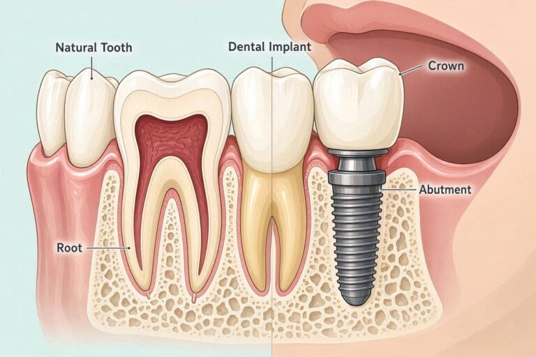
To understand why an implant will not move in an MRI, you must first understand what it is made of. The vast majority of dental implants placed worldwide are fabricated from commercially pure titanium or an alloy of titanium, aluminum, and vanadium (Ti-6Al-4V). Some modern implants use zirconia, a white ceramic material that is completely metal-free. The choice of titanium is not arbitrary; it is one of the most biocompatible materials known to science, and its magnetic properties are precisely what make it safe for imaging.
Ferromagnetism is the property that causes certain metals—iron, nickel, and cobalt—to be strongly attracted to a magnet. A ferromagnetic object in an MRI suite becomes a projectile, flying toward the bore of the magnet with lethal force. Titanium is paramagnetic. This is a much weaker, non-hazardous form of magnetism. A paramagnetic material does not retain magnetization when removed from the field. In practical terms, the massive magnetic field of a 1.5 Tesla or 3.0 Tesla MRI scanner exerts virtually zero attractive force on a titanium dental implant. Your implant is far more securely anchored by the bone it has fused to (osseointegration) than by any external force the MRI could apply. There has never been a reported case of a properly osseointegrated dental implant being dislodged by an MRI.
The other metallic concern in the mouth is not the implant fixture itself, but the restorations placed on top of it. The abutment, which connects the implant to the crown, is usually titanium or zirconia and is non-ferromagnetic. The crown itself may be made of porcelain fused to a metal alloy. Older metal alloys used in dentistry, particularly those with high nickel content, can be ferromagnetic. If your crown was made decades ago, or if you have a removable partial denture with metal clasps, these may contain magnetic metals. It is not the implant that is the primary concern; it is the unknown dental work that could cause a problem. This is why full disclosure of all your dental history is essential.
Some other dental devices are genuinely dangerous in an MRI. Aneurysm clips, some older pacemakers, and certain cochlear implants are absolute contraindications. A dental implant, however, is classified as an MR Conditional device. This means it is safe under specific conditions. The condition, in this case, is simply being embedded in the bone. The MR technologist will still follow standard safety screening protocols, and you will be asked to remove any removable dental appliances, such as dentures or retainers with metal components, before entering the scan room.
The Real Issue: Image Artifact and Distortion
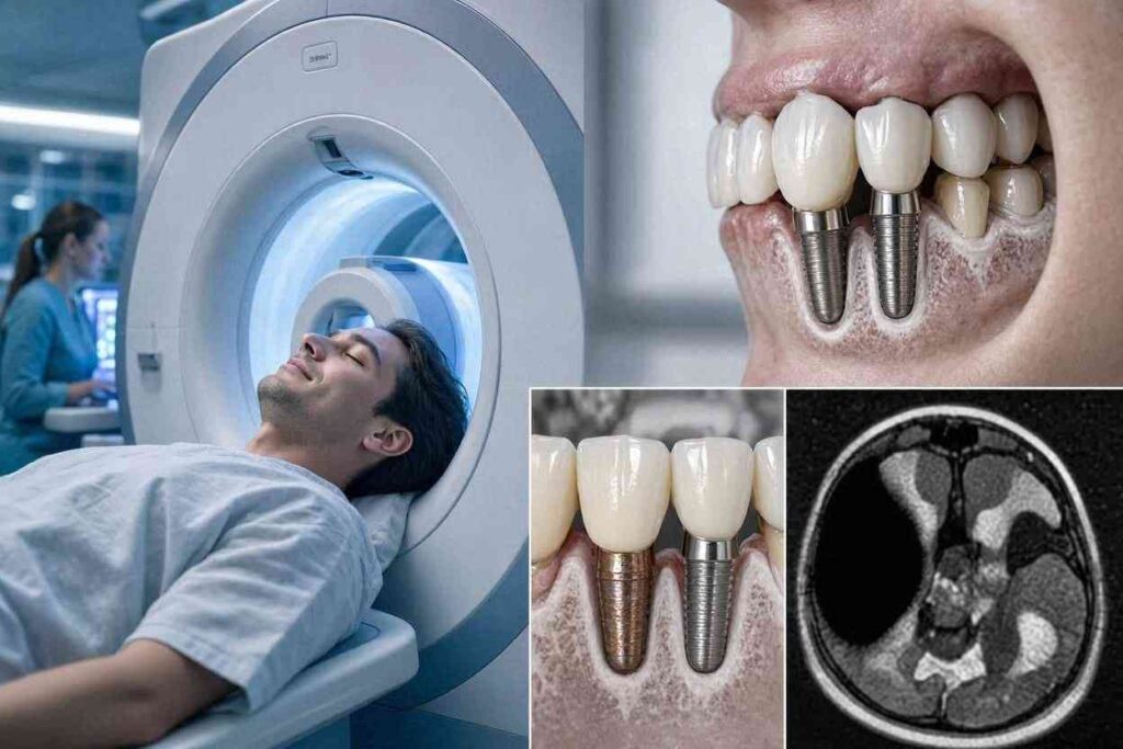
The safety of the implant is established. The diagnostic quality of the MRI image is a different conversation entirely. This is where the rubber meets the road for your radiologist. A metal object inside the body’s tissues disrupts the homogeneity of the magnetic field that the MRI machine so carefully creates. This disruption leads to a signal void and geometric distortion on the resulting image. In the simplest terms, the implant creates a black hole on the image, surrounded by a halo of warped, blurry anatomy.
The size of this artifact depends on several factors. The first is the material of the implant. Titanium causes less artifact than stainless steel, but it still causes significant distortion. Zirconia implants are virtually artifact-free because zirconia is a ceramic, not a metal. The second factor is the size of the implant. A single standard-diameter implant (about 4 millimeters) will create a small area of distortion. A full-arch set of metal implants, or implants with large metal superstructures like a cast metal bar, will cause a massive artifact. The third factor is the MRI sequence parameters. Radiologists have special metal artifact reduction sequences, known as MARS, which use specific software and scan settings to minimize the distortion.
Why does this matter to you? If the MRI is of your knee or your lower back, the artifact from your jaw implants is completely irrelevant. The metal is outside the field of view of the scan. If the MRI is of your brain, the implants in your lower jaw are far enough away from the brain parenchyma that they rarely cause diagnostic problems. The artifact might slightly distort the base of the skull or the very bottom of the temporal lobes, but the rest of the brain will be imaged clearly. However, if the MRI is of your cervical spine, your temporomandibular joints, or your face, the implant artifact becomes a major problem. A scan of the cervical spine can be rendered non-diagnostic if a mouth full of metal implants throws a massive artifact over the C1-C3 vertebrae. In these cases, the radiologist must be informed in advance to employ the MARS sequences, or an alternative imaging modality, such as a CT scan, might be necessary.
Preparing for Your MRI: The Safety Checklist
When you schedule your MRI, the imaging center will ask you a series of safety screening questions. You must not be vague. “Do you have any metal in your body?” is the trigger question. Your answer must be a clear “Yes, I have dental implants.” State how many, where they are located, and approximately when they were placed. Also, disclose any other dental work, such as braces, permanent retainers, or old metal fillings. Amalgam fillings are made of a silver-mercury-tin-copper alloy. They are not ferromagnetic, but they do contain metal and can cause small artifacts. They are not a safety hazard.
If you have any removable dental work, take it out. This includes partial dentures with metal clasps, full dentures with a metal palate, or removable retainers. These items are not integrated into your body, and if one contained a ferromagnetic component, it could move. Always err on the side of caution and leave them in your locker.
You will be asked to change into a hospital gown. This is not just for hygiene; it ensures no metallic items are hidden in your pockets. The technologist will run through the checklist again. Be patient. This repetitive screening is a safety protocol designed to prevent a catastrophic accident. Once you are on the table and positioned, the technologist will place a coil—a special antenna—around the body part being imaged. If your scan is a brain scan, the coil will cradle your head. The implants will be inside or near the coil. Again, this is safe. The coil is a receiver, not a magnet.
During the scan, you will hear loud, rhythmic banging and knocking. This is the sound of the gradient coils switching on and off. You may feel a very subtle vibration or a gentle warmth in the area of the implant. This is a normal phenomenon caused by the radiofrequency pulses inducing tiny electrical currents in the metal. It is not painful and is not a sign that anything is wrong. If you feel any discomfort, you have a call button to alert the technologist, who will stop the scan immediately.
The “Tattoo” and “Permanent Makeup” Connection
A related issue that often surprises patients is the interaction between MRI and cosmetic tattoos, including permanent lip liner, eyeliner, and microbladed eyebrows. Some tattoo pigments, especially darker colors, contain iron oxide. The MRI’s radiofrequency field can heat these particles, causing a painful burning sensation. This is a separate risk from dental implants, but if you are having a brain or facial MRI and have permanent makeup, you must disclose it. The technologist can place a cold, damp cloth over the tattooed area to act as a heat sink. The presence of dental implants does not increase the risk of a tattoo burn, but both are part of the same comprehensive safety screening.
The Impact on Different Types of MRI Scans
The clinical consequences of implant artifact vary dramatically depending on the body part being scanned. Let us walk through the most common MRI scans and how your dental implants will affect each.
Brain MRI
For a standard brain MRI ordered to investigate headaches, dizziness, or to rule out multiple sclerosis, dental implants are usually a non-issue. The implants in the mandible and maxilla are inferior to the brain. The artifact may obscure the very bottom of the temporal lobes and the sella turcica (where the pituitary gland sits). If the clinical question is about a pituitary tumor, the radiologist might need a CT scan to supplement the MRI. For the vast majority of brain pathologies, the images will be diagnostic.
Cervical Spine MRI
This is the most problematic area. The cervical spine runs from the base of the skull down to the shoulders. The top of the cervical spine is directly adjacent to the back of the jaw. If you have multiple implants in the posterior mandible or maxilla, the artifact can completely wipe out the signal from the C1, C2, and sometimes C3 vertebrae. This is a critical area for injuries and degenerative disc disease. If your doctor needs to visualize the craniocervical junction, you must inform them of your implants so they can order the MARS protocol or a CT myelogram instead of a standard MRI.
Temporomandibular Joint (TMJ) MRI
This is a specific scan ordered to evaluate the jaw joint, often for pain, clicking, or locking. Dental implants, even a single one, will create an artifact that severely degrades the image of the TMJ on that side. A TMJ MRI in a patient with dental implants is often not a useful test. The surgeon might order a CBCT or a conventional CT scan to evaluate the bony structures of the joint.
Body MRI (Abdomen, Pelvis, Spine, Knee)
Dental implants have absolutely no effect on scans of the body below the neck. The metal is too far away from the imaging field to cause any artifact. You can proceed with your lumbar spine, knee, or liver MRI without any concern about your dental work.
| MRI Scan Type | Impact of Dental Implants | Recommendation |
|---|---|---|
| Brain | Minimal; may obscure base of brain | Safe to proceed; inform technologist |
| Cervical Spine | Potentially severe; may obscure C1-C2 | Request MARS protocol; consider CT |
| TMJ | Severe; renders scan non-diagnostic | Avoid MRI; use CBCT or CT |
| Lumbar Spine/Knee/Body | None | Safe to proceed without concern |
This table is a practical guide for your conversation with your referring physician. If you have implants and your doctor is ordering a neck or jaw scan, raise this issue proactively. The doctor can then consult with the radiologist to determine the best imaging strategy.
CT Scans vs. MRI with Dental Implants
Patients often ask if a CT scan is a safer or better alternative to an MRI when they have dental implants. The answer depends entirely on the clinical question. A CT scan uses ionizing radiation (X-rays) to create images. Metal causes a different type of artifact on a CT scan, known as beam hardening artifact. This appears as bright white streaks radiating from the metal. Modern CT scanners with iterative reconstruction software can dramatically reduce this artifact, often making the bony anatomy around the implant much more visible on a CT than on an MRI.
For evaluating the bone itself—such as a fracture, a bone tumor, or the precise anatomy of the cervical vertebrae—a CT scan is often the superior choice in a patient with dental implants. For evaluating soft tissues—such as the spinal cord, the brain, the intervertebral discs, or a suspected tumor inside the jaw—MRI is superior, and the artifacts must be managed with MARS sequences.
The two modalities are complementary. It is not that one is “safe” and the other is “dangerous.” Both are safe. The implant does not move in an MRI, and the radiation dose from a focused CT is acceptable. The decision is based on which image will give the surgeon or the physician the best information to make a diagnosis.
Conclusion
Having an MRI with dental implants is unequivocally safe; the titanium or zirconia fixtures are non-ferromagnetic and firmly anchored in the bone, posing zero risk of movement or heating that would cause injury. The real clinical challenge is not safety but the potential for image artifact, which can distort the anatomy of the brain, cervical spine, or jaw and must be managed by informing the radiology team in advance. By always disclosing your dental implants during the screening process and discussing the specific body part being imaged with your doctor, you ensure the safest possible experience and the highest quality diagnostic outcome.
FAQ
Can a 3 Tesla MRI pull out a dental implant?
No. The 3 Tesla magnet, while powerful, exerts no clinically significant attractive force on titanium or zirconia dental implants. The implant is more secure in the bone than any magnetic force that could be applied to it.
Will my mouth get hot during the MRI because of the implants?
You may feel a very mild, brief warmth around the implants due to radiofrequency-induced currents. This is normal, not dangerous, and will not burn you. If you feel any actual pain, squeeze the alert ball and the scan will stop.
Are zirconia implants better than titanium for MRI imaging?
Yes. Zirconia implants cause virtually no artifact on an MRI, producing a much cleaner image with no distortion around the implant site. Titanium implants will create a signal void artifact.
Do I need to take antibiotics before an MRI if I have dental implants?
No. The current guidelines from the American Heart Association and the American Dental Association do not recommend prophylactic antibiotics for dental implants before an MRI. Prophylaxis is only for invasive dental procedures that cause bleeding.
What if I don’t know what metal my dental implant is made of?
The overwhelming majority of implants placed globally are titanium or titanium alloy. You can reasonably assume this. However, you must inform the MRI technologist that you do not know the exact metal. They will use the standard screening protocols to ensure safety.
Additional Resources
For further information on MRI safety and metallic implants, please refer to the American College of Radiology’s resources: https://www.acr.org/