What Is An Echo Scan For The Dentist?
An echo scan in a dental context is not a single, universally defined procedure with a CPT code like a medical cardiac echocardiogram. The term refers to the application of diagnostic ultrasound imaging within the dental and maxillofacial region. A dentist uses an echo scan, or ultrasound, to visualize soft tissue structures, salivary glands, lymph nodes, and vascular blood flow in real-time, without exposing the patient to ionizing radiation. This technology brings a non-invasive, dynamic diagnostic window to the oral and facial soft tissues that conventional dental X-rays and CBCT scans cannot fully provide.
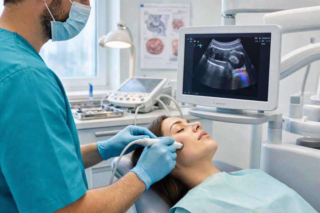
The Physics of Diagnostic Dental Ultrasound
Medical ultrasound, the technology behind an echo scan, functions on the principle of sound wave reflection. A handheld transducer, or probe, emits high-frequency sound waves, typically in the range of 5 to 18 megahertz for superficial head and neck structures. These sound waves travel through the soft tissues. When they encounter a boundary between two tissues of different acoustic impedance, such as the border between muscle and a fluid-filled cyst, a portion of the sound energy reflects back to the transducer.
The transducer detects these returning echoes and the ultrasound processing unit instantaneously calculates the depth and intensity of each reflection based on the time delay and the strength of the returning signal. This data constructs a real-time, two-dimensional grayscale image on the monitor. Blood flow can be visualized and measured using Doppler ultrasound, which detects the frequency shift of sound waves reflected off moving red blood cells. The entire process is radiation-free, painless, and provides a dynamic moving image, not a static snapshot.
The Major Clinical Applications in Dentistry
Dental echo scanning is a specialized tool most commonly used by oral and maxillofacial radiologists, oral surgeons, and specialists in oral medicine. It is not a routine screening tool in general dental practice, but its role is expanding significantly as the technology becomes more portable and affordable. The applications are specific and highly clinically valuable.
Salivary Gland Imaging and Pathology
The salivary glands are ideally suited for ultrasound examination because of their superficial location. The parotid glands, lying just in front of the ears and behind the angle of the jaw, and the submandibular glands, nestled under the lower border of the jaw, are easily accessible to an ultrasound probe. An echo scan is the first-line imaging modality for evaluating a swollen or painful salivary gland.
The scan can immediately differentiate a solid tumor, such as a pleomorphic adenoma, from a fluid-filled cyst. It can detect a salivary stone, or sialolith, which appears as a bright, hyperechoic structure with a characteristic acoustic shadow behind it. It can visualize the dilated, inflamed duct proximal to the stone. The scan also assesses the texture of the gland parenchyma, revealing the hypoechoic changes of chronic inflammation in conditions like Sjögren’s syndrome. Critically, ultrasound guides the real-time aspiration biopsy of any suspicious mass, ensuring the needle tip samples the exact target tissue.
Lymph Node Evaluation and Staging
The head and neck region contains a rich, complex network of lymph nodes that drain the oral cavity, pharynx, and face. When a patient presents with a suspicious oral lesion, an unexplained neck lump, or a known oral cancer, an echo scan of the cervical lymph nodes is an essential part of the clinical workup. The ultrasound reveals the size, shape, border, and internal architecture of the lymph nodes.
A reactive, benign lymph node from an infection is typically oval with a distinct, fatty hilum at its center. A malignant, metastatic lymph node tends to become more rounded, loses its central hilum, and may show areas of internal necrosis or abnormal peripheral blood flow on Doppler. These sonographic features directly inform the clinical staging of oral cancer, guide the decision to perform a fine-needle aspiration biopsy, and influence the extent of the planned neck dissection surgery.
Soft Tissue Masses and Cystic Lesions
The oral and perioral soft tissues can develop a wide variety of lumps and bumps, including lipomas, fibromas, hemangiomas, ranulas, and mucoceles. An echo scan provides immediate, high-resolution information about the nature of these lesions. It can confirm whether a blue, dome-shaped swelling on the lower lip is a fluid-filled mucocele or a vascular lesion.
A ranula, which is a mucus escape cyst in the floor of the mouth, can be visualized plunging through the mylohyoid muscle into the neck. The relationship of the cyst to the sublingual gland and the surrounding muscles is mapped in real time. This diagnostic clarity is obtained instantly, in the office, without any radiation dose and at a significantly lower cost than an MRI.
Tongue and Oropharyngeal Imaging
High-resolution, small-footprint intraoral ultrasound probes are a newer development. These allow the dentist or radiologist to scan the tongue directly. The primary application is the assessment of the depth of invasion of a squamous cell carcinoma of the tongue. The thickness of the tumor, measured precisely on ultrasound, is a critical factor in predicting the risk of lymph node metastasis and planning the surgical margins. The scan can also visualize tongue base lesions and assess the lateral pharyngeal wall.
Temporomandibular Joint Effusion
While CBCT is the standard for imaging the bony components of the temporomandibular joint, an echo scan can detect an effusion, or fluid accumulation, within the joint capsule. This appears as a dark, hypoechoic area within the joint space. A joint effusion is a sign of active inflammation, and its presence can help guide the decision to perform an arthrocentesis, a procedure where the joint is flushed to remove the inflammatory mediators.
The Advantages and Limitations of Dental Ultrasound
The dental echo scan occupies a specific and powerful niche. Its primary advantages are the absence of ionizing radiation, real-time dynamic imaging, relatively low cost, and the ability to correlate the image directly with the patient’s physical anatomy during the exam. It is painless and non-invasive.
Its limitations are equally clear. Ultrasound does not penetrate bone well, so it cannot image structures deep to the cortical bone of the mandible or maxilla. It cannot diagnose caries or periapical bone lesions, which remain the domain of X-rays and CBCT. The quality of the scan is highly operator-dependent. A skilled, experienced clinician-sonographer is essential for obtaining and interpreting diagnostically valid images. It is not a replacement for CT or MRI but a complementary, problem-solving tool that, in specific soft tissue questions, provides the definitive answer most elegantly.
Conclusion
An echo scan in dentistry is a diagnostic ultrasound examination used to visualize the soft tissues of the head and neck, including the salivary glands, lymph nodes, and oral soft tissue masses, without radiation. It provides real-time, dynamic imaging that instantly differentiates solid from cystic lesions, guides biopsies, evaluates the depth of oral tumor invasion, and assesses lymph node architecture in cancer staging. While it cannot see through bone or diagnose tooth decay, it is an indispensable, specialized tool that integrates seamlessly with three-dimensional X-ray imaging to provide a complete diagnostic picture.
Frequently Asked Questions
Q: Is an echo scan the same as the ultrasound used during pregnancy?
A: Yes, it is the exact same technology and physical principle. The difference is that a dental ultrasound uses a smaller, higher-frequency probe designed for the very superficial anatomy of the face and neck, providing much higher resolution detail than an abdominal obstetric scan.
Q: Can my general dentist perform an echo scan on my jaw joint for TMJ pain?
A: Some general dentists who have received specific training and certification in head and neck ultrasound may offer this service. However, a diagnostic ultrasound is most often performed and formally interpreted by an oral and maxillofacial radiologist or a surgeon in their practice.
Q: Is a dental echo scan painful or risky?
A: It is completely painless and non-invasive. A small amount of gel is applied to the skin to allow the sound waves to transmit, and the smooth probe is gently moved over the area of interest. There are no known biological risks at the diagnostic ultrasound intensities used.
Additional Resource
For professional standards and guidelines on the safe and effective use of diagnostic imaging, including ultrasound, in dental and maxillofacial practice, the leading specialty academy is the definitive source.
- American Academy of Oral and Maxillofacial Radiology: aaomr.org