Why Does A Dental Implant Fail?
You trusted the process. You underwent surgery. You endured the healing period. You paid a significant sum of money. You received your new crown and for a while, everything was perfect. Then something changed. The implant feels loose. Your dentist uses words you dread: “The implant is failing.” Your heart sinks. How could this happen? You did everything you were told. You thought implants were supposed to last a lifetime. Why does a dental implant fail?
Dental implant failure is a devastating outcome for both patient and clinician. Understanding why implants fail is the first step toward preventing failure and making informed decisions if you face this situation. This comprehensive guide will dissect the two categories of implant failure, early failure before the crown is placed, and late failure after the implant has been in function. We will explore every major risk factor, from surgical technique to systemic disease to patient habits. By the end of this article, you will understand the complex interplay of factors that determine implant survival.
Defining Implant Failure
Implant failure is not a single event. It is a process that culminates in the loss of the implant. Clinically, an implant is considered a failure if it is mobile, if it has lost a significant amount of supporting bone beyond a threshold defined by success criteria, if it causes persistent pain or infection that cannot be resolved, or if it fractures. An implant that has failed must be removed. The distinction between a failing implant, which is compromised but may be salvageable, and a failed implant, which is beyond saving, is important. This article focuses on the factors that lead to the final common pathway of implant loss.
Early Implant Failure
Early failure occurs before the implant is restored with a crown, or shortly after crown delivery, typically within the first few months to a year. The primary mechanism of early failure is the failure of osseointegration. The bone does not successfully fuse with the implant surface. The implant is essentially a foreign body that the bone has rejected or to which it has failed to attach.
Surgical Trauma and Overheating the Bone
The most common cause of early failure is excessive surgical trauma, particularly overheating the bone during the drilling process. The osteotomy, the hole drilled into the bone for the implant, must be prepared with sharp, well-cooled drills. The drills generate frictional heat. If the bone temperature exceeds 47 degrees Celsius, or about 117 degrees Fahrenheit, for more than one minute, the bone cells die. This is called thermal necrosis. A zone of dead bone surrounds the implant. The body cannot heal. The implant fails to integrate.
Preventing thermal necrosis is a fundamental surgical skill. The surgeon uses copious irrigation with sterile saline, a progressive series of sharp drills, and an intermittent drilling technique, lifting the drill to allow coolant to reach the depth. A surgeon in a hurry who forces a dull drill into dense bone is courting disaster. This is entirely preventable.
Contamination of the Implant Surface
The titanium surface of a dental implant is engineered to be osteoconductive, meaning it encourages bone cells to attach and grow. This surface is chemically active. If the surface is contaminated during surgery, the bone cells will not attach.
Contamination can occur if the implant touches the patient’s lips, cheek, or saliva before placement. It can occur if the surgeon’s gloves have powder or contaminants on them. It can occur if the surgical site is not adequately debrided of inflammatory tissue from a failing tooth. A strict sterile protocol is essential. The implant should only touch the bone, the sterile irrigation fluid, and the sterile titanium instruments. Even a brief contact with a non-sterile surface can be enough to compromise osseointegration.
Poor Bone Quality and Quantity
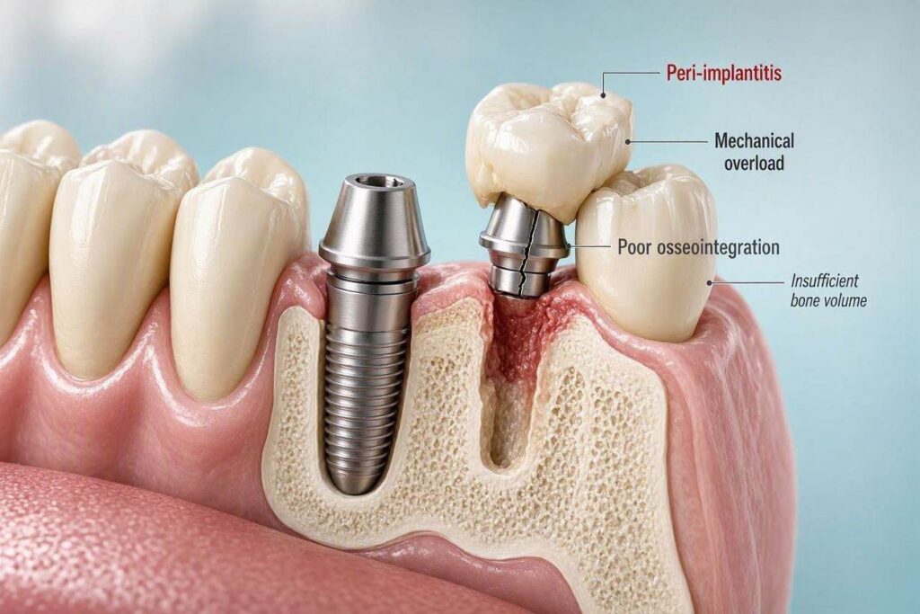
The patient’s bone must be adequate in both quantity and quality to support the implant. If there is insufficient bone volume, the implant may not be fully encased in bone. Part of the implant may be exposed, or it may be placed in a graft that has not fully matured. If the bone quality is poor, which is often described as Type IV bone, the bone is very soft and spongy with a thin cortical plate. This is commonly found in the posterior maxilla. The initial stability of the implant, called primary stability, is difficult to achieve in soft bone. Without primary stability, the implant micromoves during healing. Micromotion prevents osseointegration. A fibrous capsule forms around the implant instead of bone. The implant fails.
Perforation of the Buccal Plate
If the implant is placed too far toward the cheek, it can perforate the thin buccal plate of bone. The implant tip or threads protrude through the bone, exposed to the soft tissue. Bone cannot grow where there is no bone. The exposed surface invites soft tissue ingrowth and infection.
Infection at the Surgical Site
A preexisting infection at the site, such as a chronic abscess from a failing tooth, can overwhelm the healing process. The bacteria colonize the implant surface before the bone cells can reach it. Adequate debridement of the socket after extraction and a period of healing before implant placement reduce this risk.
Late Implant Failure
Late failure occurs after the implant has successfully osseointegrated and has been restored with a crown. The implant was successful, but then it begins to fail, often years later. The primary mechanisms are peri-implantitis and occlusal overload.
Peri-Implantitis
As discussed in a previous article, peri-implantitis is a bacterial infection that causes inflammation and progressive bone loss around a functioning implant. The bacteria accumulate in the sulcus, form a biofilm, and trigger a host inflammatory response. The bone is destroyed. The implant loses its support. Peri-implantitis is the leading cause of late implant failure. The risk factors for peri-implantitis, poor oral hygiene, history of periodontitis, smoking, uncontrolled diabetes, and excess cement, are the same risk factors for late failure. Meticulous long-term maintenance is the only defense.
Occlusal Overload
An implant can fail if it is subjected to excessive, uncontrolled biting forces. Unlike a natural tooth with a periodontal ligament that absorbs shock, an implant is rigidly fixed in the bone. Excessive forces are transmitted directly to the bone-implant interface, causing microfractures in the bone and eventual loss of integration.
Overload can occur due to a poorly designed bite, a premature contact, or a cantilevered prosthesis. It is most common in patients who grind or clench their teeth severely, a condition called bruxism. Bruxism generates forces that far exceed normal chewing forces. Without a protective nightguard, these forces can cause the implant to fracture, the abutment screw to break, or the supporting bone to resorb.
Implant Fracture
The implant fixture itself can fracture, although this is rare with modern titanium implants. Fracture is usually due to a combination of heavy occlusal forces and a design weakness, such as a narrow-diameter implant used in a molar site, or an implant with an internal connection that has been over-stressed. A fractured implant is a catastrophic failure. The remaining piece of the implant must be surgically removed, which can be a challenging procedure.
Abutment Screw Loosening and Fracture
The abutment screw, the tiny screw that connects the abutment to the implant, is the mechanical weak link in the system. It is designed to be the point of failure to protect the implant itself from fracture. Repeated loosening of the abutment screw is a sign of excessive forces or a poor fit. Eventually, the screw can fatigue and fracture. The broken screw fragment is difficult to retrieve. If it cannot be retrieved, the implant may need to be removed.
Systemic Risk Factors for Implant Failure
Certain patient-specific factors significantly increase the risk of implant failure.
Smoking
Smoking is one of the strongest and most consistent risk factors for implant failure. Smokers have significantly higher failure rates than non-smokers. Nicotine is a potent vasoconstrictor. It reduces blood flow to the gums and bone, impairing healing and the delivery of immune cells and nutrients. Smoking impairs the function of neutrophils and other immune cells. The heat and chemical toxins in cigarette smoke also directly irritate the oral tissues and alter the bacterial flora. Smokers are at much higher risk for both early failure, due to poor healing, and late failure, due to peri-implantitis. Many implant surgeons will not place implants in heavy smokers unless they commit to smoking cessation.
Uncontrolled Diabetes
Diabetes mellitus, particularly when poorly controlled with elevated hemoglobin A1c levels, impairs wound healing, reduces resistance to infection, and alters bone metabolism. Microvascular disease in diabetics reduces blood flow to the bone, similar to smoking. Implant success rates are lower in diabetic patients with poor glycemic control. Well-controlled diabetics have success rates approaching those of non-diabetics.
Immunocompromised States
Patients taking immunosuppressive medications, such as corticosteroids or chemotherapy agents, patients with HIV/AIDS with low CD4 counts, and patients undergoing cancer treatment have compromised healing and increased infection risk. Implant placement in these patients requires careful medical consultation and a case-by-case risk assessment.
Osteoporosis and Bisphosphonates
Osteoporosis itself is not a direct contraindication to implants, but the quality and density of the bone may be reduced. The greater concern is patients taking bisphosphonate medications, such as alendronate (Fosamax) or zoledronic acid (Reclast). Bisphosphonates are used to treat osteoporosis and certain cancers. They profoundly suppress bone turnover. This can lead to a devastating complication called medication-related osteonecrosis of the jaw (MRONJ), where the jaw bone dies and fails to heal after trauma, such as an extraction or implant surgery. Patients on intravenous bisphosphonates for cancer are at very high risk and are generally not candidates for implant surgery. Patients on oral bisphosphonates for osteoporosis are at lower risk, but this risk increases with the duration of therapy.
Radiation Therapy to the Jaw
Patients who have received high-dose radiation therapy to the head and neck region for cancer treatment are at risk for osteoradionecrosis, a condition similar to MRONJ. The irradiated bone has a permanently compromised blood supply and reduced healing capacity. Implant placement in irradiated bone carries a higher risk of failure and of triggering bone death. Hyperbaric oxygen therapy may be used before and after surgery to improve healing.
The Surgeon Factor
The skill and experience of the implant surgeon are critical variables. Implant surgery is a technically demanding procedure. The surgeon must accurately assess the bone, plan the implant position using 3D imaging, execute the surgery atraumatically, and manage any complications that arise. A poorly placed implant, in the wrong position, at the wrong angle, or with damage to adjacent structures, is set up for failure. Choose a surgeon who places implants regularly, not occasionally, and who uses modern diagnostic and surgical techniques.
Summary of Key Causes of Implant Failure
- Early Failure: Caused by failure of osseointegration due to surgical overheating, contamination, poor bone quality, lack of primary stability, or preexisting infection.
- Late Failure: Caused primarily by peri-implantitis, a bacterial infection leading to bone loss, and by occlusal overload from excessive biting forces or bruxism.
- Patient Factors: Smoking, uncontrolled diabetes, immunocompromised states, bisphosphonate use, and prior radiation therapy significantly increase failure risk.
- Mechanical Factors: Implant fracture and abutment screw fracture, often related to bruxism or poor prosthetic design, can cause catastrophic failure.
Conclusion
Dental implants fail because of a disruption in the delicate biological equilibrium between the implant, the bone, and the oral environment. Early failure is primarily a surgical and healing problem, where the bone fails to integrate with the implant. Late failure is primarily a maintenance and biomechanical problem, where infection or excessive forces destroy the bone that supports a once-successful implant. Understanding your personal risk factors, choosing a skilled surgeon, committing to meticulous oral hygiene, and attending regular maintenance visits are the pillars of preventing implant failure. If an implant does fail, prompt removal, site rehabilitation with bone grafting, and re-evaluation of the risk factors can often pave the way for a successful replacement implant.
Frequently Asked Questions (FAQ)
1. What are the first signs of a failing dental implant?
Signs include mobility, a loose feeling, bleeding or pus around the implant, a dull ache or pain when biting, gum recession exposing the implant threads, and swelling or redness of the gum.
2. Can a failed dental implant be replaced?
Yes, in most cases. The failed implant is removed, the site is debrided, and a bone graft is often placed. After healing, typically three to six months, a new implant can be placed.
3. What percentage of dental implants fail?
The overall survival rate for dental implants is high, around 95% to 98% over 10 years. However, failure rates are higher in smokers, patients with uncontrolled diabetes, and in areas of poor bone quality.
4. Is implant failure the dentist’s fault?
Not always. Failure can be due to patient factors like smoking or poor hygiene, or biological factors like poor bone quality. However, poor surgical technique, such as overheating the bone or placing the implant in a bad position, is a preventable cause.
5. Does a failed implant hurt when it is removed?
The removal procedure is done under local anesthesia. Post-operative pain is usually manageable and similar to the original implant surgery.
6. Can an X-ray show if an implant is failing?
An X-ray can show bone loss around the implant, which is a key sign of failing. However, an X-ray alone cannot diagnose failure; clinical signs like mobility must also be present.
7. How can I prevent my implant from failing?
Do not smoke. Maintain meticulous oral hygiene. See your dentist for regular implant maintenance visits. Wear a nightguard if you grind or clench your teeth. Control your diabetes if you are diabetic.
Additional Resource
For more information on dental implant care and complications, visit the American Academy of Implant Dentistry:
American Academy of Implant Dentistry – Patient Information