What Is A Ct Scan For Dental Implants?
You are on the path to a dental implant. Your dentist or oral surgeon has told you that you need a “CT scan” or a “3D scan” before the surgery can be planned. You may be familiar with medical CT scans from hospitals. You lie on a table, and a large, donut-shaped machine passes over you. The dental world uses a different, smaller, and more focused technology. The question is straightforward: what is this scan, why is it absolutely necessary, and what does it reveal?
This article is a deep dive into the Cone Beam Computed Tomography (CBCT) scan, the gold standard diagnostic image for dental implant planning. We will explain how it differs from a medical CT and a routine dental panoramic X-ray, what the surgeon sees in the detailed slices of the image, and why this scan is the single most important safety tool in modern implant dentistry.
Beyond the Flat Image: The Limitation of 2D X-Rays
For decades, dentists relied on two-dimensional X-rays to see the jawbone. A panoramic X-ray, the one where the machine rotates around your head, produces a single, flat, wide image showing all the teeth, the jaws, the sinuses, and the temporomandibular joints. It is a useful screening tool, a map of the general terrain.
However, a panoramic X-ray has a fundamental limitation. It is a two-dimensional shadow of three-dimensional structures. It compresses all the anatomy into one flat plane. It shows the height of the bone, but it cannot show the width or the depth. It cannot tell the surgeon how thick the bone is on the cheek side or the tongue side. Critically, it cannot show the precise, three-dimensional location of the inferior alveolar nerve canal, the vital sensory nerve that runs through the lower jaw, relative to a proposed implant site. A 2D X-ray shows the nerve as a dark band, but it cannot reveal whether the nerve is positioned toward the cheek, toward the tongue, or directly in the path of the planned implant. To place an implant safely, the surgeon needs to see the bone as a volume, not a silhouette.
The 3D Solution: Cone Beam Computed Tomography (CBCT)
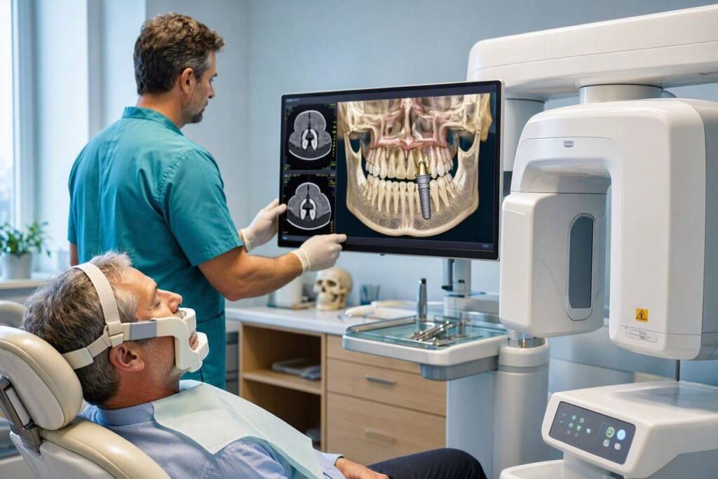
The Cone Beam CT, or CBCT, is the technology that solved this problem for dentistry. It is a specific type of CT scanner designed for the maxillofacial region. Unlike a medical CT scanner, which uses a fan-shaped beam and requires the patient to lie flat on a table that moves through a large gantry, a dental CBCT scanner is typically smaller, and the patient sits or stands upright.
The “cone beam” refers to the shape of the X-ray beam, which is a cone, not a fan. The machine rotates once around the patient’s head, capturing hundreds of individual two-dimensional projection images in a single 360-degree sweep. A powerful computer reconstructs these hundreds of images into a single, three-dimensional, volumetric digital model of the entire maxillofacial skeleton. This scan can be viewed as a solid 3D model, or it can be sliced into thin, cross-sectional views, like looking at individual pages of a book, each page representing a millimeter-thick slice of the jaw.
The radiation dose from a focused dental CBCT scan is significantly lower than a standard medical head CT, but it is higher than a single panoramic X-ray. The dose is clinically justified by the life-changing diagnostic information it provides. The scan is fast, non-invasive, and comfortable. You simply sit still while the machine rotates around you.
What the Surgeon Sees: The Anatomy in Three Dimensions
When the surgeon loads the CBCT scan onto their planning software, they can manipulate the 3D model of your jaw in any direction. They can rotate it, zoom in, and measure distances with sub-millimeter accuracy. This is what they are looking for, and this is why the scan is non-negotiable.
The Bone Volume and Density
The scan reveals the exact height, width, and three-dimensional shape of the residual alveolar ridge. The surgeon can measure the available bone in millimeters at every point along the ridge. They can assess the density of the bone, which affects the implant’s primary stability. A site that looks adequate on a 2D panoramic X-ray may be revealed by the CBCT to be a knife-edge ridge, far too thin to house an implant without a bone graft.
The Vital Structures: The Nerve and the Sinus
In the lower jaw, the surgeon traces the path of the inferior alveolar nerve canal in 3D. They can see its exact buccal-lingual position. They can measure the distance from the crest of the ridge to the roof of the nerve canal. This measurement directly determines the maximum safe length of the implant. The implant must end at least one to two millimeters above the nerve to prevent permanent nerve damage. A 2D X-ray cannot provide this safety margin with certainty.
In the upper jaw, the surgeon visualizes the maxillary sinus floor. They can see the exact shape of the sinus cavity, identify any thickening of the sinus membrane (indicative of chronic sinusitis), and measure the available vertical bone beneath the sinus. This determines whether a sinus lift is necessary.
The Pathology
The CBCT can reveal silent, asymptomatic pathology that a routine 2D X-ray might miss. A residual cyst from an old extraction, a retained root tip, an impacted tooth, or a pathological lesion in the bone will be clearly visible. The surgeon can diagnose and plan for the management of these unexpected findings before the day of surgery, avoiding an intra-operative surprise.
The Implant Simulation and Surgical Guide Design
The most powerful use of the CBCT is the virtual implant simulation. Using specialized software, the surgeon and restorative dentist virtually place the implant into the 3D bone model. They select the exact implant system, diameter, and length. They position it in the prosthetically ideal location, angling it perfectly to support the future crown. They can verify that the implant sits safely away from the nerve and the sinus, with adequate bone surrounding it on all sides. This virtual surgery is performed on the computer before a single scalpel touches your gum. From this virtual plan, the precision surgical guide is designed and 3D-printed.
The Legal Standard of Care: The use of CBCT imaging for dental implant planning has become the standard of care in modern implant dentistry. A surgeon who places an implant based only on a 2D panoramic X-ray, and who subsequently injures a nerve or perforates a sinus, is practicing below the accepted standard. The CBCT is not an optional upgrade. It is the diagnostic foundation of safe, predictable, and prosthetically driven implant surgery.
The Patient Experience: What to Expect During the Scan
Having a dental CBCT scan is a simple, quick, and comfortable experience. You will be asked to remove any metal jewelry, eyeglasses, and hearing aids from the head and neck region, as metal can create artifacts on the image. You may wear a lead apron as a precaution. You will be positioned in the machine, either standing or seated, with your chin resting on a support and your forehead against a band. Small, soft positioning guides may be placed in your ears to ensure your head is perfectly aligned.
The operator will instruct you to remain completely still and not to swallow for the short duration of the scan, which is typically 20 to 40 seconds. The machine will rotate silently around your head. There is no sensation, no noise, and no claustrophobia. It is over in less than a minute. The scan data is then processed, and the detailed images are ready for your surgeon to review within minutes.
Conclusion
A CT scan for dental implants, specifically a Cone Beam CT, is a three-dimensional X-ray that provides a precise, volumetric digital model of your jaws. It is the essential diagnostic tool that allows the implant surgeon to measure bone volume and density, precisely locate the vital nerves and sinuses, identify hidden pathology, and perform a complete virtual implant simulation before the day of surgery. It is the map that guides a safe, predictable, and prosthetically driven implant outcome.
Frequently Asked Questions
Is a dental CT scan safe? How much radiation is it?
A dental CBCT scan delivers a very low, focused dose of radiation, significantly less than a medical CT scan of the head. The dose is equivalent to a few days of natural background radiation. The diagnostic benefit of avoiding a nerve injury or a failed implant far outweighs the minimal radiation risk.
Do I need a referral for a dental CT scan?
A dental CBCT scan is typically ordered by your implant surgeon or restorative dentist as part of the implant treatment planning process. You do not need a separate referral from a physician. The scan is performed in the dental office if they have the equipment, or at a dedicated dental imaging center.
Will my dental insurance cover the cost of the CBCT scan?
Many comprehensive dental PPO plans cover a portion of the cost of a CBCT scan when it is deemed medically necessary for implant planning. It is typically classified under a specific radiology code. Check with your insurance provider. The scan fee often ranges from $250 to $600, and may be credited toward the total implant fee by some practices.
Additional Resource:
For a detailed, patient-friendly explanation of Cone Beam CT imaging and its role in dental diagnosis, visit the American Academy of Oral and Maxillofacial Radiology: https://www.aaomr.org/patient-information/